Offer
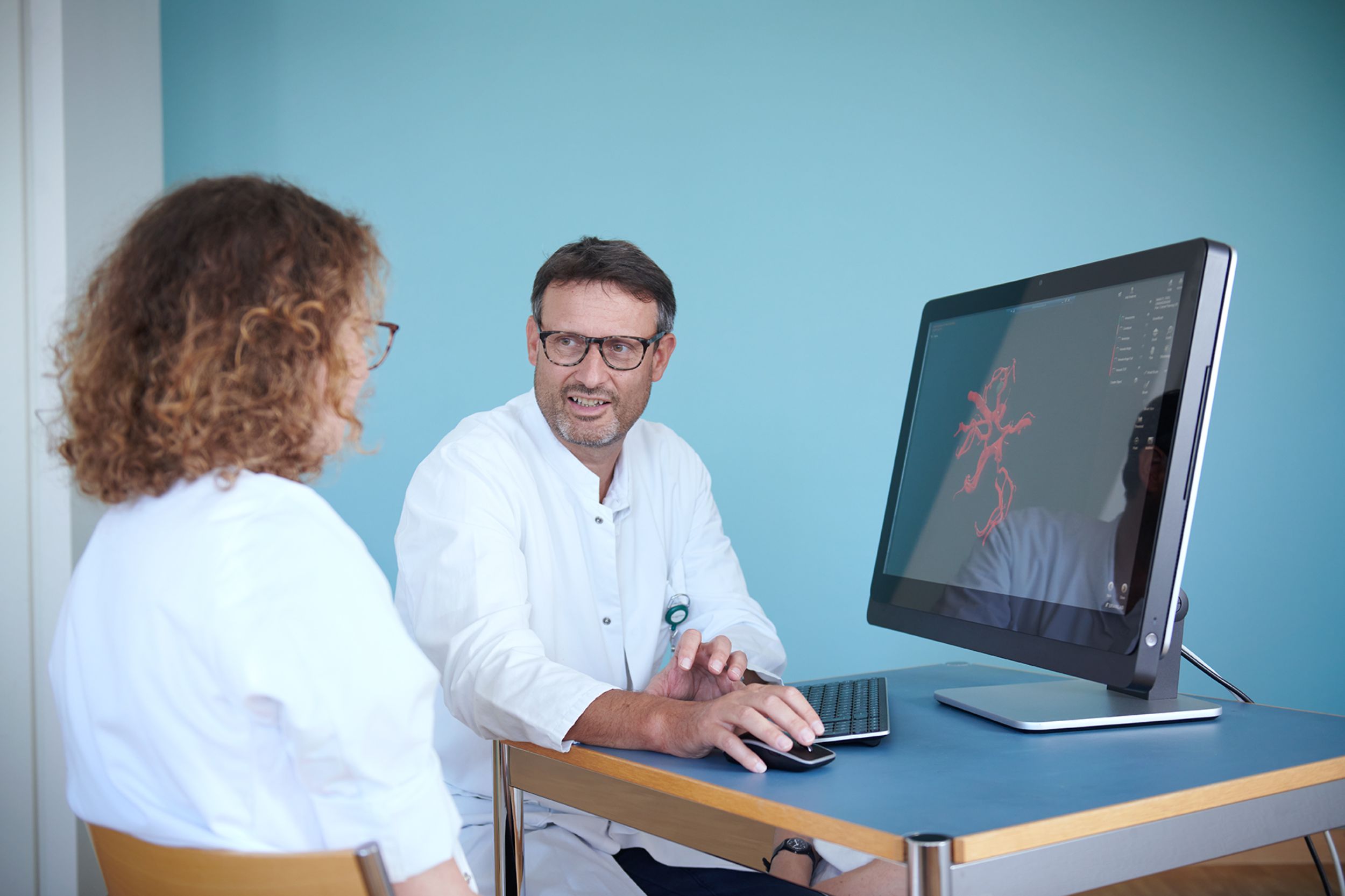
The brain and spinal cord, also known as the "central nervous system", control almost all functions in the body. Injuries or diseases of the central nervous system are very diverse and lead to a wide range of symptoms. This is why a precise and rapid diagnosis and treatment of these diseases by specialized personnel is important and central.
Our clinic offers a wide range of neurosurgical examinations and interventions on the brain. Injuries and diseases of the spine and spinal cord are treated by the Spinal Surgery Clinic.
Range of services
Brain tumors
As a matter of principle, our Brain Tumor Center strives to treat our patients in an interdisciplinary consensus. For you as a patient, this means that your "case" will be discussed in our interdisciplinary brain tumor conference and that you as a patient will be offered a treatment strategy recommended by all relevant specialist disciplines. In addition to neurosurgery, these specialist disciplines include oncology, radiation oncology, neuroradiology, neuropathology and neurology.
The tumors treated in this consultation include
- Low-grade and high-grade gliomas
- meningiomas
- Brain metastases
- Malformation tumors
- Lymphomas of the central nervous system
- Tumors of the spinal cord and spinal nerves
The central element of the consultation is always the personal discussion. During this consultation, all available diagnostic and therapeutic measures are presented and discussed, including the objectives, risks and prospects of success. During this consultation, it is often very helpful to bring along relatives or other people close to the patient, as the situation can then be viewed from another perspective. The special consultation for brain tumors is also available to obtain second opinions.


Skull base & pituitary glands
A specialized team of neurosurgeons, hormone specialists, ear, nose and throat specialists, ophthalmologists and radiation specialists is available to advise patients with diseases of the skull base. In interdisciplinary conferences for brain tumors (brain tumor board) and specialized for tumors of the pituitary gland (pituitary board), an individually adapted treatment strategy is determined for the affected patients.
Consultations take place in the skull base consultation hour in relation to
- Pituitary tumors
- Skull base meningiomas
- Vestibular schwannomas (acoustic neuromas)
- Rarer skull base tumors
- Spontaneous cerebrospinal fluid leakage (rhinoliquorrhea)
The patient is advised on appropriate follow-up, minimally invasive transnasal endoscopic and microtechnical transcranial operations at the base of the skull (olfactory groove, pituitary lodge, cerebellopontine angle, foramen magnum) and adjuvant procedures such as stereotactic radiosurgery and radiotherapy.
Prof. Daniel Bodmer
Chefarzt
HNO Leitung
Neurovascular consultation
A specialized team of neurosurgeons, neuroradiologists and neurologists is available to advise patients with cerebrovascular diseases. In the interdisciplinary conference for neurovascular diseases (Neurovascular Board), an individually tailored treatment strategy is determined for affected patients.
Consultations take place in the neurovascular consultation hour in relation to
- Aneurysms
- Cerebral vascular malformations (arteriovenous malformation, dural arteriovenous fistula)
- cavernomas
- strokes
The patient is advised on appropriate follow-up, surgical and/or endovascular treatment of cerebrovascular disease and other procedures such as stereotactic radiosurgery.
Prof. Raphael Guzman
Chefarzt und Klinikleiter
Neurochirurgie / Pädiatrische Neurochirurgie
Leiter Innovations-Focus Pädiatrische Neurochirurgie
Consultation hours for functional neurosurgery and hydrocephalus
For the consultation of patients with functional disorders and hydrocephalus, individually adapted treatment strategies for affected patients are determined in this consultation.
In the functional neurosurgery and hydrocephalus consultation hour, consultations are held in relation to
- Parkinson's disease, essential tremor, dystonia and other movement disorders for which treatment with deep brain stimulation (DBS) is an option
- typically as part of an overall evaluation prior to DBS by the multidisciplinary functional stereotaxy team USB
- Trigeminal neuralgia (as a disease in its own right or as a symptom of an underlying disorder such as multiple sclerosis)
- Other chronic pain syndromes that may be treatable by neurosurgery
- hydrocephalus
- in particular so-called normal pressure hydrocephalus or hydrocephalus malresorptivus
The patient will be advised on the individual indication and selection of possible neurosurgical treatment options.
Pediatric neurosurgery consultation (UKBB)
A neurosurgical team specializing in children is available to advise children with brain or spinal cord diseases. An individually tailored treatment strategy for the affected patients is determined at interdisciplinary conferences.
In the pediatric neurosurgery consultation hour, consultations are held in relation to
- Cranial neurosurgery (head)
- Brain tumors
- Cranial calvaria tumors
- Hydrocephalus (hydrocephalus)
- Intracranial cysts (neuro-endoscopy)
- Intracranial pressure problems (pseudotumor cerebri etc.)
- Surgical treatment of epilepsy (open surgical treatment/vagus nerve stimulator implantation)
- Cerebrovascular diseases (vascular diseases of the brain)
- Craniocerebral trauma
- Skull and facial malformations (craniosynostosis)
- Encephalocele
Pediatric spinal neurosurgery (spinal column and spinal cord):
- Spinal cord tumors
- Spinal dysraphia (myelomeningocele, spinal lipomas, filum terminale lipomas, spinal cord clefts)
- Surgical treatment of cerebral palsy/spasticity (single-level selective dorsal rhizotomy, baclofen pump/catheter)
- Intervertebral disc problems (herniated disc, narrowing of the spinal canal with slipped vertebrae)
- Chiari malformation and other malformations of the cranio-cervical junction
Our interdisciplinary consultation hours:
- Craniofacial consultation (for craniosynostosis together with our oral surgeons)
- Tonus consultation (for spasticity together with our neuropaediatricians and neuroorthopaedists)
- MMC consultation (for dysraphia together with our neuropaediatricians, pediatric nephrologists, neuroorthopaedists and pediatric urologists)
- Pediatric neuro-oncology consultation (for brain and spinal cord tumors together with our pediatric neuro-oncologists, neuropathologists, pediatric radiologists and neuropaediatricians)
Parents and child(ren) are advised on sensible follow-up checks and surgical treatments for pediatric neurosurgical diseases and other procedures.
Further information on the Center for Pediatric Neurosurgery (USB-UKBB) can be found on our website and on the UKBB website.

Prof. Raphael Guzman
Chefarzt und Klinikleiter
Neurochirurgie / Pädiatrische Neurochirurgie
Leiter Innovations-Focus Pädiatrische Neurochirurgie

Dr. Maria Lucia Licci
Oberärztin
Neurochirurgie / Pädiatrische Neurochirurgie
Pädiatrische Neurochirurgie
Private consultation
Patients with supplementary insurance generally have the option of choosing their neurosurgeon in the inpatient area. Accordingly, we also offer outpatients the opportunity to consult with the head physician or the head physician of their choice, especially if surgical treatment is being considered.
You can navigate to more detailed descriptions of our range of treatments below.


Prof. Raphael Guzman
Chefarzt und Klinikleiter
Neurochirurgie / Pädiatrische Neurochirurgie
Leiter Innovations-Focus Pädiatrische Neurochirurgie


Below you can navigate to more detailed descriptions of our range of treatments.
Vascular neurosurgery
Vascular neurosurgery is a subspecialization of neurosurgery that deals with and treats diseases of cerebral vessels. It primarily involves the treatment of patients with vascular malformations, stroke and carotid stenosis.
The treatment of vascular diseases at the University Hospital Basel is carried out in an interdisciplinary setting, consisting of highly specialized vascular neurosurgeons, interventional neuroradiologists and vascular neurologists. Our institution is the only one in Switzerland that has dual-trained neurosurgeons (neurosurgery and interventional neuroradiology). This leads to ideal collaboration between the two disciplines in everyday practice and in our new, state-of-the-art hybrid operating theater, where surgical and endovascular procedures can be performed simultaneously.
Patients with vascular neurosurgical diseases are discussed weekly at our interdisciplinary stroke colloquium and our specialists determine the best treatment option for each patient. Patients with stroke are assessed and treated efficiently, quickly and professionally by our highly specialized Stroke Unit.
Aneurysm
An aneurysm is a sac-like protrusion caused by a weakness in the vessel wall. This weakness can be congenital or acquired in connection with certain secondary diseases (e.g. high blood pressure).
The thin-skinned wall of an aneurysm can rupture and cause life-threatening bleeding in the brain (subarachnoid hemorrhage), which can lead to symptoms such as severe, sudden headaches, neurological deficits such as paralysis or even coma. Aneurysms that do not burst typically cause no symptoms and are usually discovered by chance during imaging of the head (incidentally discovered aneurysms).
A ruptured aneurysm with subarachnoid hemorrhage is a major emergency situation and treatment involves closure of the aneurysm and long-term intensive medical and neurosurgical follow-up. As a rule, patients require an inpatient stay in a neurorehabilitation center after acute treatment. An incidental aneurysm is not considered an emergency situation and, depending on the size, configuration and location of the aneurysm, it must either be closed or closely monitored.
Treatment of the aneurysm can either be closed (endovascular), using special microcatheters and platinum coils (coiling) or open by means of surgery (clipping). The respective therapy is determined individually for each patient by our highly specialized interdisciplinary neurovascular team, consisting of vascular neurosurgeons and interventional neuroradiologists.
Thanks to our new hybrid operating theater, which is equipped with endovascular instruments, we offer vascular care at the highest and most modern level. We can complete operations on cerebral vessels (such as clipping) and immediately check the endovascular result or even combine open, surgical and endovascular procedures and perform them in the operating theater in the same session.
Cerebral vascular malformations
Tangles and short circuits of vessels are known as arteriovenous malformations (AVM). In the brain, such malformations are often diagnosed in childhood and young adulthood (20 to 30-year-olds). As a rule, these malformations are congenital, and in around 40 percent of cases a cerebral hemorrhage is the first sign of the disease. However, AVM can also manifest itself through paralysis, epileptic seizures or headaches.
Our highly specialized interdisciplinary neurovascular team, consisting of vascular neurosurgeons and interventional neuroradiologists, determines the treatment of an AVM individually for each patient. Treatment may include surgical, endovascular or radiotherapeutic measures or a combination of all these techniques.
Thanks to our new hybrid operating theater, which is equipped with endovascular instruments, we offer vascular care at the highest and most modern level. We can complete operations on cerebral vessels (such as AVM occlusion) and immediately check the result endovascularly or even combine open, surgical and endovascular procedures and perform them in the same session in the operating theater.
Cavernomas
Cavernomas (cavernous hemangiomas) are usually acquired vascular malformations of the brain. They occur 80 percent in the cerebrum and 15 percent in the cerebellum or brain stem.
Cavernomas can remain asymptomatic for life and in such cases are often discovered by chance during imaging of the head. Symptomatic cavernomas often cause epileptic seizures, but can also lead to chronic headaches, cerebral hemorrhages and focal neurological deficits (paralysis, speech disorders and others).
Cavernomas do not always need to be treated immediately, but if discovered, they must be monitored closely. In the event of a cerebral hemorrhage, correlating symptoms or growth of the cavernoma, treatment should be considered. The treatment is usually surgical removal of the cavernoma. However, this is not always possible and depends on the location in the brain.
Stroke (stroke)
A stroke occurs when the oxygen supply to the brain is interrupted. There is a reduced blood supply to one or more areas of the brain, which leads to the death of brain tissue and cells. This is known as a stroke.
Causes of a stroke can be acute vascular occlusion due to the spread of blood clots (embolisms), vascular constriction (stenosis), infection, increased intracranial pressure or cerebral hemorrhage. However, a cerebral infarction can also lead to a secondary hemorrhage in the brain, which is known as a hemorrhagic stroke.
Depending on the area and side of the brain affected, various symptoms occur, such as paralysis, speech disorders, impaired vision and even a coma. In the event of a stroke, the time to treatment plays a major role. To ensure that the process from assessment to treatment is carried out as quickly as possible, stroke patients are treated in our Stroke Unit. This unit is specially set up for strokes and is staffed by a team of neurosurgeons, neurologists and neuroradiologists. As a result, the patient receives the best possible holistic and fastest clarification and treatment.
The treatment of a stroke is primarily carried out endovascularly, with the aim of reopening the blocked or narrowed vessels. In certain cases, a neurosurgical operation is necessary, particularly in the case of severe swelling of the brain caused by the stroke (so-called malignant infarction) or in the case of cerebral hemorrhage, which leads to increased intracranial pressure. In such cases, part of the skull is removed in order to reduce the pressure in the brain and thus enable the patient to survive, or if the cerebral hemorrhage needs to be surgically removed.
Carotid stenosis
Carotid stenosis is a narrowing of the carotid artery. Depending on the degree of narrowing, there is an increased risk of a stroke. If the degree of stenosis is over 80 percent, the annual risk of a stroke is around 3 percent.
The cause of carotid stenosis is usually vascular calcification in the arteries supplying the brain. This calcification is caused by smoking, high cholesterol levels, high blood pressure, old age or diabetes, among other things.
Carotid stenosis can be asymptomatic and in such cases is discovered either by chance or during routine examinations. Symptomatic carotid stenosis often leads to short-term neurological deficits ("minor stroke", TIA or PRIND), such as paralysis, speech or visual disturbances, but these are considered warning symptoms before a stroke.
The aim of treatment if carotid stenosis is detected is to prevent a stroke. Depending on the degree of stenosis and symptoms, conservative (drug) therapy or invasive (surgical or endovascular) therapy is available. Surgical therapy consists of removing the calcifications (plaques) in the area of the stenosis and is called thrombendarterectomy. Endovascular therapy involves widening the vessel using a balloon with or without the insertion of a metal mesh tube (stent), which keeps the vessel open. Both interventions are carried out with the help of neuromonitoring, whereby brain wave measurements are taken during the operation to ensure maximum safety for the patient. All patients with carotid stenosis are discussed on an interdisciplinary basis at our weekly stroke colloquium, where our specialists determine the best treatment options for each patient individually.
Craniocerebral injuries (traumatic brain injury)
A traumatic brain injury describes an injury to the skull and brain structures, usually caused by an accident or assault. The extent of a traumatic brain injury can vary greatly - from a laceration without permanent consequences to a severe brain injury or hemorrhage with permanent serious damage to the brain and body.
Mild traumatic brain injuries can usually be treated conservatively with pain-relieving medication and rest. Severe brain injuries may require life-saving emergency surgery followed by intensive medical treatment and long-term neurorehabilitation in a specialized clinic.
Cerebral hemorrhages with craniocerebral injuries
Various types of cerebral hemorrhage can occur as a result of a traumatic brain injury. Some require life-saving emergency surgery and others can be treated without surgery. Bleeding after a traumatic brain injury is classified according to its anatomical localization.
Epidural hemorrhage: This is a hemorrhage between the bone and the hard meninges (dura). This results in the rupture of an arterial blood vessel, usually with rapid development of a hemorrhage. An increase in the size of the hemorrhage causes increasing pressure on the brain, which can lead to symptoms. Typical is a brief loss of consciousness at the scene of the accident with rapid recovery afterwards. If the hemorrhage increases in the course of the procedure, it can lead to renewed clouding and even coma. For these reasons, epidural hemorrhage often requires immediate neurosurgical emergency surgery. During the operation, the bone is opened, the hemorrhage removed, the bleeding vessel closed and the bone reinserted. The prognosis is usually very favorable as there are no concomitant brain injuries.
Acute subdural hemorrhage: This is a fresh hemorrhage between the hard meninges (dura) and the surface of the brain. The source of bleeding is often veins that run between the dura and the brain and rupture in the event of trauma. Depending on the time course, a distinction is made between acute (fresh) and chronic (old) subdural hemorrhages. In acute subdural hemorrhage, there is usually rapid bleeding with pressure on the brain, so that surgical relief is often necessary. As there are often accompanying brain injuries in these cases, the prognosis is poorer and the mortality rate is relatively high (30 to 80 percent). The prognosis is also heavily dependent on the patient's age. The only treatment for symptomatic patients is immediate surgery to remove the hemorrhage. This operation often involves removing a large piece of the skull. In most cases, this piece of bone is not reinserted at the end of the operation, as the accompanying brain injuries often cause severe swelling of the brain during the course of the operation.
Chronic subdural hemorrhage: This is an old hemorrhage between the hard meninges (dura) and the brain, which accumulates slowly over several weeks and can often only lead to symptoms later on. This hemorrhage often occurs in older patients after minor injuries. Typical symptoms are headaches, personality changes, gait disorders and paralysis. Symptomatic chronic hemorrhages are treated by surgery using two drill holes in the skull, flushing out the blood and leaving a drain in place for 24 to 48 hours. The prognosis is favorable, although around 10 percent of patients suffer a relapse of the bleeding (recurrence), which usually leads to a repeat operation.
Intracerebral hemorrhage (contusion hemorrhage): These are hemorrhages directly in the brain tissue. The symptoms depend on the size and localization of the hemorrhage. These hemorrhages are usually treated without surgery, although large space-occupying hemorrhages may require surgery. The prognosis depends on the age of the patient and the location of the hemorrhage.
Shear injuries of the brain
Shearing injuries of the brain, also known as "diffuse axonal injuries" or "shearing injuries", are a special form of traumatic brain injury with a relatively poor prognosis. Shearing injuries often occur when the head hits soft surfaces, such as the paneling of a car, as the force on such surfaces acts evenly on the brain. The injury pattern is therefore diffuse, i.e. distributed throughout the brain, but the individual areas of the brain are only slightly injured and barely visible. Nevertheless, shearing injuries often lead to severe disabilities, as the entire brain is affected by the injury. Surgical treatment is only necessary in the case of generalized brain swelling (cerebral edema), but shearing injuries cannot be treated surgically.
Skull calvaria fracture (fracture)
Skull fractures, also known as skull fractures, are not uncommon in the context of craniocerebral injuries. A distinction is made between dislocated (i.e. displaced), non-dislocated, comminuted and wedged fractures. A distinction is also made between open and closed fractures. A special subtype of skull fracture is the skull base fracture.
Most skull fractures do not require surgical treatment and can be treated conservatively with pain medication and rest. Fractures that lead to an injury to the dura mater or brain tissue, are wedged or dislocated, usually require surgical treatment. Fractures of the base of the skull can also usually be treated conservatively. In rare cases, cerebrospinal fluid (CSF) may leak out, in which case it must be surgically sealed.
The prognosis for skull fractures depends on the accompanying brain injury. If there is no brain injury, the prognosis is very good.
Brain tumors
Abnormal tissue that grows through uncontrolled cell division and damages the surrounding tissue is called a tumor. Brain tumors can develop from brain tissue (gliomas), meninges (meningiomas), or as offshoots of tumors in other organs in the body (brain metastases).
The symptoms of a brain tumor are very variable and depend on the location of the tumor and its size. Brain tumors often cause few symptoms, especially tumors that grow slowly and in non-functional areas of the brain. Possible signs of a brain tumor include headaches, nausea, recurrent vomiting, changes in personality, paralysis and epileptic seizures.
Brain tumors are usually treated surgically, with total removal usually being the goal. Depending on the location of the tumor and its size, complete resection is sometimes not possible or advisable. In such cases, a (needle) biopsy is recommended.
All patients with brain tumors are discussed as part of our interdisciplinary tumor board and an individual therapy is determined for each patient. Further aftercare takes place within the framework of our Brain Tumor Center, consisting of neurosurgeons, neuro-oncologists and neuro-radiotherapists.
The neurosurgery clinic at the University Hospital Basel is very involved in glioma research. In addition to his clinical work, Prof. Luigi Mariani heads a brain tumor research laboratory (Brain Tumor Biology Laboratory), where, among other things, the biology of gliomas is studied and possible innovative therapies are developed (e.g. immunotherapies).
Prof. Dr. med. Dr. sc. nat. Gregor Hutter was awarded a professorship by the Swiss National Science Foundation (SNSF) and is researching novel therapies for malignant brain tumors with his group.
Film on the subject of awake craniotomy:
Gliomas
Gliomas are the most common brain tumors in adults. They develop from the supporting tissue (glial cells) of the nervous system. Some types of glioma can occur in childhood, others only develop in adulthood. In principle, a distinction is made between low-grade gliomas and high-grade gliomas.
Low grade gliomas:These are referred to as grade II gliomas according to the WHO classification. They usually occur in younger to middle-aged patients (typically between 30 and 45 years of age), but can also occur in childhood. Low-grade tumors typically grow slowly and therefore lead to relatively late and only mild symptoms. The slow growth allows the brain to shift important functions from areas of the brain affected by tumors to other, healthy regions of the brain. Because these tumors grow slowly and lead to mild symptoms, they are usually discovered relatively late, when they have already grown to a considerable size. Typically, epileptic seizures can be the first indication of low-grade gliomas. Other non-specific symptoms such as headaches, mood changes, fatigue, dizziness, unsteady gait, nausea and vomiting are also possible. An MRI examination leads to a suspected diagnosis and referral to neurosurgery.
Treatment usually consists of surgical removal, firstly to reduce the pressure in the brain caused by the tumor and secondly to obtain an accurate histological diagnosis. The more radical the resection, the higher the survival rate after resection. The aim of the operation is therefore to remove the tumor as completely as possible without impairing the body's functions (speech, motor function, etc.). Depending on the location and size of the tumor, only a partial resection or biopsy (small tissue removal) may be performed. As these types of tumors often grow adjacent to or within important brain centers (e.g. speech center, movement center, etc.), new modern tools are used, such as the surgical microscope, neuronavigation and intraoperative monitoring. In the case of tumors near the speech or visual center, the operation must be performed while the patient is awake, as these functions cannot be monitored while the patient is asleep.
At the University Hospital Basel, such operations are regularly carried out while the patient is awake, and a highly specialized team of neurosurgeons, neuroanesthetists and neuropsychologists is specially trained for these procedures. As a rule, chemotherapy and radiotherapy are not used in the first instance for low-grade gliomas, but only if they develop into high-grade gliomas.
The prognosis for low-grade gliomas depends on various factors. We know from studies that the extent of resection is the most important prognostic factor for long-term survival. Age under 50 years, small tumor volume and few symptoms also have a favorable influence on the prognosis. In addition, molecular genetic factors (e.g. mutation of the p53 gene, mutation 1p19qLOH, IDH mutation, etc.) of the tumor influence the prognosis.
High-grade gliomas: The two most common brain tumors are glioblastoma (WHO grade IV) and anaplastic astrocytoma (WHO grade III). These tumors usually occur between the ages of 50 and 60 and arise from the supporting cells of the brain (so-called glia).
Depending on the location of the tumor in the brain, different symptoms arise. A tumor near the speech center leads to speech problems, a tumor near the movement center can lead to loss of strength. In addition, non-specific symptoms such as headaches, epileptic seizures, altered consciousness, fatigue, dizziness, unsteady gait, nausea and vomiting can occur. These tumors typically lead to swelling of the surrounding brain tissue, which intensifies the symptoms. For this reason, drug therapy with steroids (fortecortin), which is often administered in advance, alleviates the symptoms.
New modern aids are also used in these operations, such as the surgical microscope, neuronavigation and intraoperative monitoring, as well as the modern technology of blue light fluorescence technology (5-ALA), which helps us to reliably identify and remove the tumor. Before the operation, modern imaging techniques such as functional MRI (fMRI), which provides valuable information on the localization of speech function and movement centers, or MRI-based fibre tracking, which shows the exact course of important nerve pathways deep in the brain, are used as required. After surgical tumor resection, additional drug treatment (chemotherapy) and radiation (radiotherapy) is usually initiated. After the final histological assessment of the removed tissue, the individual treatment plan is determined at the weekly tumor board of our Brain Tumor Center.
High-grade gliomas: High-grade gliomas: The two most common brain tumors are glioblastoma (WHO grade IV) and anaplastic astrocytoma (WHO grade III). These tumors usually occur between the ages of 50 and 60 and arise from the supporting cells of the brain (so-called glia).
Depending on the location of the tumor in the brain, different symptoms arise. A tumor near the speech center leads to speech problems, a tumor near the movement center can lead to loss of strength. In addition, non-specific symptoms such as headaches, epileptic seizures, altered consciousness, fatigue, dizziness, unsteady gait, nausea and vomiting can occur. These tumors typically lead to swelling of the surrounding brain tissue, which intensifies the symptoms. For this reason, drug therapy with steroids (fortecortin), which is often administered in advance, alleviates the symptoms.
New modern aids are also used in these operations, such as the surgical microscope, neuronavigation and intraoperative monitoring, as well as modern blue light fluorescence technology (5-ALA), which helps us to reliably identify and remove the tumor. Before the operation, modern imaging techniques such as functional MRI (fMRI), which provides valuable information on the localization of speech function and movement centers, or MRI-based fibre tracking, which shows the exact course of important nerve pathways deep in the brain, are used as required. After surgical tumor resection, additional drug treatment (chemotherapy) and radiation (radiotherapy) is usually initiated. After the final histological assessment of the removed tissue, the individual treatment plan is determined as part of the weekly tumor board at our Brain Tumor Center.
Meningiomas
Meningiomas are generally benign, slow-growing tumors that arise from a layer (arachnoid) of the meninges. According to the WHO classification, meningiomas are divided into three stages (WHO grade I-III). WHO grade I meningiomas are benign, occur most frequently and have a low growth rate and a low risk of recurrence. Higher grade meningiomas (WHO grade III) are more aggressive, but occur much less frequently and have an increased growth rate, a tendency to infiltrate the surrounding tissue and a higher risk of recurrence. WHO grade II meningiomas are regarded as an intermediate stage between grade I and grade III meningiomas.
Meningiomas occur more frequently from the age of 50, with women being affected about twice as often as men.
The cause of meningiomas is still unknown. Ionizing radiation is considered a risk factor; according to studies, people who have been exposed to a particularly high dose of radiation have a significantly higher risk (around 6 to 10 times higher) of developing a meningioma. An additional risk factor for developing meningiomas is the genetic disease neurofibromatosis type 2.
The treatment of symptomatic meningiomas or meningiomas that increase in size over time consists of surgical removal as completely as possible. Small and non-symptomatic meningiomas do not need to be operated on, but require an annual imaging check, as they can increase in size over time. In most cases, complete removal of the tumor means a cure for the patient. In cases where complete removal is not possible (for example, if the tumor adheres to important vessels or areas of the brain), radiotherapy offers a good alternative to reduce the growth of the residual tumor.
Film on the topic of preoperative consultation - meningioma:
Vestibular schwannomas (acoustic neuromas)
Vestibular schwannoma is a benign tumor of the Schwann cells, the supporting cells of the vestibular nerves. The symptoms usually manifest themselves as unilateral hearing loss, hearing noise (tinnitus) and/or balance disorders. Despite their benign nature, these tumors pose a challenge for treatment, depending on their size, due to their delicate localization with a very close relationship to the auditory nerve, the facial nerve (facial nerve) and often the brain stem.
A wait-and-see approach is possible for small tumors. Growth is monitored in regular radiological examinations using MRI. However, the chances of a hearing-preserving operation are significantly better in the early stages.
Surgical removal with monitoring of cranial nerve function by neuromonitoring is suitable for tumors of any size. Small tumors can also be treated with targeted radiation (radiosurgery). In most cases, further growth of the tumor can be prevented in this way. The results and complication rates of surgery and radiosurgery are largely comparable. The aim of surgical treatment is to remove as much of the tumor as possible while preserving cranial nerve functions (hearing, facial mobility, balance and coordination). Facial paralysis is a severe impairment for the patient, so preserving the facial nerve is a top priority during treatment. Hearing preservation on the affected side is possible in about half of the cases, but this depends very much on the size and nature of the tumor. Permanent balance disorders and other complications are very rare and can usually be treated well.
Pituitary tumors
The pituitary gland is a pea-sized gland that hangs on the lower surface of our brain (pituitary gland) and controls the entire hormonal balance of our body. Approximately one in five tumors of the brain is a pituitary tumor. The most common tumors of the pituitary gland are adenomas, which are practically always benign. Depending on their size, a distinction is made between small microadenomas (<10 mm) and larger macroadenomas (>10 mm). Other rarer tumors of the pituitary region are craniopharyngiomas, metastases, cysts (so-called Rathke cysts) or germinomas.
Tumors of the pituitary gland cause symptoms due to the loss of hormonal functions, pressure on the optic nerves or an overproduction of hormones. As a rule, pituitary adenomas are removed through the nose using a minimally invasive transsphenoidal approach. Some pituitary adenomas are treated initially or permanently with tablets (typically prolactinomas). Other tumors require surgical treatment, which is performed endoscopically assisted by an interdisciplinary team (neurosurgery and ENT). In rare cases, the tumor must be removed via an access through the skull (craniotomy).
All patients with pituitary tumors are discussed in our interdisciplinary pituitary board, consisting of ear, nose and throat specialists, endocrinologists, ophthalmologists and radiation specialists, and an individual therapy is determined for each patient.
Brain metastases
Brain metastases are offshoots of cancer cells from other organs. They are the most common tumors in the brain. Up to a third of all patients with advanced cancer develop brain metastases. Metastases can occur in the cerebrum, cerebellum or meninges. In up to 50 percent of cases, a single (singular) metastasis occurs, but multiple metastases can also occur. The most common metastases in the brain originate from breast or lung cancer.
The symptoms vary depending on the location of the metastasis in the brain. They can trigger non-specific symptoms of increased intracranial pressure (headaches, nausea, vomiting) or symptoms such as epileptic seizures, paralysis or speech disorders. Sometimes brain metastases are discovered by chance without symptoms occurring. As a rule, metastases are diagnosed by means of an MRI of the head. If another cancer in the body (primary tumor) is not known, a CT scan of the chest and abdomen is used to actively search for a primary tumor.
The treatment options for a brain metastasis are surgical removal or radiosurgical irradiation. As a rule, these two methods are supplemented with drug therapy (chemotherapy). The decision as to which metastasis should be operated on and which should be treated with radiosurgery is made individually at the weekly tumor board of our Brain Tumor Center.
Hydrocephalus (hydrocephalus)
Cerebrospinal fluid (CSF) surrounds the brain and spinal cord and flows through the ventricles, which are located in the brain. Up to 0.5 liters of this fluid are produced every day, which means that with an average cerebrospinal fluid content of around 150 ml, the cerebrospinal fluid is completely turned over approximately three times a day. The cerebrospinal fluid is produced in the brain in the so-called choroid plexus and flows out again via the so-called Pacchioni granulations. Overproduction or outflow disorders of cerebrospinal fluid can lead to an increased accumulation and build-up of cerebrospinal fluid in the ventricular system, which is known as hydrocephalus (hydrocephalus). Brain infections, brain tumors, aging processes, cerebral hemorrhages and malformations are typical causes of hydrocephalus. Hydrocephalus typically leads to increased pressure inside the head, which results in symptoms.
Typical symptoms of hydrocephalus are headaches, nausea, vomiting, memory disorders, gait disorders, bladder dysfunction (incontinence), visual disturbances and loss of consciousness or even coma.
The diagnosis is made by means of imaging of the head (MRI or CT) and possible measurement of intracranial pressure by means of a puncture in the back (lumbar puncture).
The aim of treatment is to restore the normal flow of cerebrospinal fluid or to create an alternative outflow. The treatment is usually surgical. In the acute situation, a so-called external ventricular drainage (EVD) is inserted, allowing the cerebrospinal fluid to drain outwards via a catheter into a system connected to it. Otherwise, if possible, the cause leading to the hydrocephalus is removed (e.g. removal of the tumor or cyst in the ventricle). If this is not possible, an alternative drainage pathway can be created using a ventriculoperitoneal shunt (VP shunt) or a neuro-endoscopic procedure (see below).
Normal pressure hydrocephalus
In normal pressure hydrocephalus, which mainly occurs in older people, there is a slow increase in the volume of cerebrospinal fluid inside the skull over a relatively long period of time. There is no or only a very slight increase in pressure, as the increase in cerebrospinal fluid volume is accompanied by a loss of brain mass; symptoms are therefore often delayed and gradual. Normal pressure hydrocephalus is most noticeable in the loss of brain function. Typical of this is the Hakim triad with small-step, broad-based gait, cognitive or memory disorders and incontinence.
As the brain's ability to absorb cerebrospinal fluid is permanently impaired, no causal treatment is possible. The treatment of choice is the implantation of a VP shunt. A silicone catheter is inserted through the skull roof into the ventricular system using anatomical landmarks or ultrasound. This catheter is connected via a subcutaneous reservoir, which later allows a puncture of the system (e.g. for cerebrospinal fluid examination), to a valve that controls the outflow of cerebrospinal fluid depending on the pressure in the head and can be regulated from the outside using a magnet.
From the valve, the catheter is guided under the skin into the abdominal cavity, where the cerebrospinal fluid is absorbed. In the event of inflammation in the abdominal cavity or adhesions due to previous abdominal surgery, there is the alternative of diverting the catheter into the heart (so-called ventriculoatrial shunt/VA shunt).
Hydrocephalus malresoptivus
In hydrocephalus malresoptivus, the resorption of cerebrospinal fluid is impaired while cerebrospinal fluid production remains unchanged. Bleeding in the brain caused by vascular diseases, vascular malformations or after craniocerebral trauma, meningitis and brain surgery can lead to hydrocephalus malresorptivus. The underlying disease increases the content of blood products and/or proteins in the cerebrospinal fluid. As a result, the outflow pathways of the cerebrospinal fluid in the ventricular system become blocked and/or sticky, leading to an imbalance between cerebrospinal fluid production and reabsorption and ultimately to hydrocephalus and increased pressure in the head. In such cases, the treatment of choice is also the insertion of a VP shunt.
Hydrocephalus occlusivus (occlusive hydrocephalus)
In occlusive hydrocephalus, the outflow of cerebrospinal fluid within the ventricles of the brain is impaired. This can be caused by tumors, scarring (sometimes also congenital) or cysts in or next to the ventricular system. An additional cause is aqueductal stenosis. In this clinical picture, the aqueduct, the narrowest point in the ventricular system, is narrowed or blocked by a membrane (which is usually congenital), which leads to a chronic build-up of cerebrospinal fluid and hydrocephalus. The treatment of occlusive hydrocephalus is varied and usually depends on the cause.
In the case of tumors or cysts, it may be possible to restore the normal flow of cerebrospinal fluid by surgically removing the tumor or cyst. If tumor or cyst removal is deemed too risky or is not possible, a detour for the cerebrospinal fluid can be created via a minimally invasive opening in the area of the ventricular system (endoscopic third ventriculostomy). In certain unclear processes, an endoscopic (camera-guided) third ventriculostomy can be performed to treat the hydrocephalus and biopsy the tumor endoscopically during the same procedure. The final option is the insertion of a VP shunt if all other options are not possible or have been exhausted.
Functional neurosurgery
Functional neurosurgery involves the surgical treatment of neurological conditions that do not - or no longer - respond adequately to purely medical therapy. These include involuntary movement disorders such as Parkinson's syndrome, tremor of other causes (such as essential tremor or tremor caused by multiple sclerosis) or dystonia. These are usually treated with deep brain stimulation (see below). Treatment of pain syndromes such as trigeminal neuralgia or chronic back pain, which are treated using spinal cord stimulators or the insertion of drug pumps in the area of the spinal cord, are also part of functional neurosurgery (performed by our colleagues in spinal surgery). Finally, the surgical treatment of epilepsy is also part of functional neurosurgery.
Trigeminal neuralgia
The fifth cranial nerve, the trigeminal nerve, provides the sensory supply to the forehead and face and the motor supply to the masticatory muscles. Trigeminal neuralgia causes pain in the sensitive supply area of the trigeminal nerve. The pain typically occurs in attacks and is often triggered by chewing or touching. The patient's quality of life is often very limited due to the high intensity of the pain. Compression of the trigeminal nerve as it exits the brain stem by a pulsating cerebral vessel (so-called neurovascular conflict) or by a tumor can be the cause of trigeminal neuralgia. On the other hand, trigeminal neuralgia can be idiopathic, i.e. without a clear cause, or as part of other nerve diseases such as multiple sclerosis.
The diagnosis is usually clinical, i.e. based on the symptoms described (without additional examinations). To clarify a possible cause (e.g. neurovascular conflict or tumor), an MRI of the head is performed, which influences the appropriate therapy. If a process in the area of the trigeminal nerve (e.g. tumor or cyst) can be seen, it will be surgically removed if possible. Otherwise, trigeminal neuralgia is primarily treated with medication (e.g. carbamazepine (Tegreteol), pregabalin (Lyrica) and/or gabapentin (Neurontin)).
If drug therapy does not lead to satisfactory pain relief, invasive/surgical treatment may be considered. These include so-called microvascular decompression of the trigeminal nerve, glycerol infiltration or thermocoagulation. Microvascular decompression is primarily performed when a microvascular conflict is detected on MRI. The trigeminal nerve is surgically exposed via an opening in the skull (craniotomy) and the vessel that is pulsating and compressing in the area of the nerve is located. The compressing vessel is then permanently separated from the nerve by a Teflon pad. Glycerol infiltration or thermocoagulation of the trigeminal nerve is performed under local anesthesia (patient is awake) in the operating theatre.
Under X-ray control, the area in which the so-called ganglion of the trigeminal nerve (ganglion gasseri) is located is located and punctured through the cheek using a needle. During glycerol infiltration, the drug glycerol is applied locally and during thermocoagulation, thermal damage is caused to the nerve through heat. This is intended to destroy the pain fibers of the nerve, which leads to pain relief. This treatment can also be repeated several times.
Deep brain stimulation (DBS)
Deep brain stimulation is used for neurological diseases such as Parkinson's disease or hemiballismus (special forms of truncal ganglion disease with violent, involuntary, skidding movements of the limbs) that cannot be treated satisfactorily with medication. The most common indication is Parkinson's syndrome. Before deep brain stimulation is carried out, our neurology colleagues try to stabilize the disease with medication. In certain cases, drug therapy is not sufficient, so deep brain stimulation is discussed. Deep brain stimulation is primarily used when Parkinson's patients have severe motor impairments or severe tremor despite good drug treatment and do not show any severe cognitive problems or behavioral abnormalities. In this case, the patient is referred to our movement disorder consultation for planning of deep brain stimulation by the treating neurologist after a thorough assessment.
Deep brain stimulation is a surgical procedure. Small microelectrodes are placed with millimeter precision in a deep brain region (subthalamic nucleus, globus pallidus internus or nucleus ventralis intermedius thalami, depending on the clinical picture and symptoms) and connected to an impulse generator, which is usually located under the collarbone. This operation is performed in a so-called stereotactic frame, which enables the electrodes to be placed in the brain with millimeter precision. Following hospitalization, all patients undergo inpatient rehabilitation, which is planned and organized in advance.
Epilepsy surgery
The uncontrolled electrical discharge from nerve cells in the brain can lead to epileptic seizures. If these seizures recur, this is epilepsy in the narrower sense. The quality of life of patients with chronic epilepsy is severely impaired, especially if the seizures occur repeatedly despite the use of various medications. Many of these patients have focal epilepsy, i.e. a specific area of the brain is "sick" and triggers the seizures. It is often possible to identify the diseased brain region and remove it surgically. The results of this surgery are very good, with up to 90 percent of operated patients becoming seizure-free. At the University Hospital Basel, we have special expertise in this cutting-edge medical field, where neurologists and neurosurgeons work very closely together.
Vagus nerve stimulator (VNS)
For epilepsy patients who do not respond adequately to drug therapy and for whom surgical treatment is not possible or has not been able to significantly reduce the seizures, implantation of a so-called vagus nerve stimulator (VNS) is an option. A battery-operated stimulation device is implanted in the area of the chest muscles and connected to the vagus nerve (10th cranial nerve) in the neck area via fine electrodes. The stimulation device regularly sends weak electrical impulses to the nerve cords of the vagus nerve leading to the brain.
The full effect of the VNS on epilepsy is not yet fully understood. Among other things, it is assumed that the activation of the vagus nerve and its nerve branches has a positive influence on various brain regions involved in the development of epilepsy. In around 30 to 50 percent of patients, a reduction in seizures of around 30-50 percent can be seen with the help of a VNS, which has a significant effect on the quality of life of these patients. However, complete freedom from seizures is rarely achieved. The operation is performed under general anesthesia, but in an outpatient setting (i.e. patients can usually return home on the same day) and has a relatively low complication rate. Recently, more and more patients suffering from depression are also being treated with VNS and the overall results are relatively promising.
Pediatric neurosurgery
The pediatric neurosurgery department at the University Children's Hospital Basel (UKBB) provides surgical care for children with brain or spinal cord diseases. Prof. Raphael Guzman and Prof. Jehuda Soleman have additional training in pediatric neurosurgery and cover the entire spectrum of neurosurgical diseases in children and adolescents. The highly specialized and experienced team has access to the latest surgical techniques and equipment.
Thanks to intensive national and international research activities, pediatric neurosurgery at the UKBB offers the latest findings and experience, which benefit children and adolescents during their treatment. In addition to optimal medical therapy, care and attention are top priorities for the treatment team. The pediatric neurosurgery department at UKBB is the first in Switzerland to cover the entire spectrum of neurosurgical diseases in children and adolescents with specially trained neurosurgeons.
Main areas of pediatric neurosurgery
Cranial neurosurgery (head):
- Brain tumors
- Cranial calvaria tumors
- Hydrocephalus (hydrocephalus)
- Intracranial cysts
- neuro-endoscopy
- intracranial pressure problems (pseudotumor cerebri etc.)
- Surgical treatment of epilepsy (open surgical treatment/vagus nerve stimulator implantation)
- Cerebrovascular diseases (vascular diseases of the brain)
- Craniocerebral trauma
- Skull and facial malformations (craniosynostosis)
- encephalocele
Pediatric spinal neurosurgery (spine and spinal cord):
- Spinal cord tumors
- Spinal dysraphia
- Myelomeningocele
- Spinal lipomas
- Filum terminale lipomas
- Surgical treatment of cerebral palsy/spasticity (single-level selective dorsal rhizotomy, baclofen pump/catheter)
- Intervertebral disc problems (herniated disc, narrowing of the spinal canal with slipped vertebrae)
- Chiari malformation and other malformations of the cranio-cervical junction
Further information on the Center for Pediatric Neurosurgery (USB-UKBB) can be found on the UKBB website.
