COVID-19 Recherche
Principaux investigateurs

Prof. Matthias Matter
Leitender Arzt und Fachbereichsleiter Molekularpathologie
Pathologie
Tel. +41 61 328 64 71

Prof. Alexandar Tzankov
Leitender Arzt und Fachbereichsleiter Histopathologie und Autopsie
Pathologie
Tel. +41 61 328 68 80
Membres du groupe


Notre science
Interactions SARS-CoV-2 avec des cellules, des tissus et des organes humains chez des patients COVID-19
Une pandémie en constante évolution, causée par le coronavirus 2 du syndrome respiratoire aigu sévère (SRAS-CoV-2) et conduisant à la maladie à coronavirus 2019 (COVID-19), a dominé le début de l'année 2020 et continue de le faire. Des cas de COVID-19 ont été décrits pour la première fois à la fin de l'année 2019, lorsqu'une série de décès liés à la pneumonie, non identifiés auparavant, sont survenus dans la province du Hubei, en Chine. COVID-19, s'est ensuite propagé à presque tous les pays avec plus d'un demi-million de décès confirmés dans le monde (https://www.worldometers.info/coronavirus/).
Bien que l'origine du virus, l'entrée cellulaire et l'épidémiologie aient été rapidement clarifiées, les observations in situ des interactions virales réelles au sein des organes et des tissus humains chez les patients souffrant de COVID-19 ont été abordées pendant longtemps au niveau des rapports de cas ou des petites séries de ≤4 cas. En effet, à la fin du mois d'avril 2020, alors que 150 000 patients étaient déjà décédés de COVID-19, seuls 16 cas d'autopsie avaient été rapportés dans la littérature évaluée par les pairs, avec neuf publications présentant des autopsies limitées, l'évaluation de la nécrose corticale post-mortem ou la collecte incisionnelle de tissus. En l'absence de données fiables sur le degré d'infection par le SRAS-CoV-2 chez les personnes décédées, diverses autorités ont découragé la réalisation d'autopsies. Cette situation, combinée à l'attitude peu enthousiaste des pathologistes, des cliniciens et des sociétés à l'égard des autopsies, a freiné les activités scientifiques visant à élucider les véritables mécanismes sous-jacents du COVID-19, ce qui semble incompréhensible compte tenu du rôle fondamental et chronophage des autopsies dans les maladies émergentes ou inconnues. Ce n'est qu'après 170.000 décès rapportés dus au COVID-19 et quatre mois de pandémie que la première série d'autopsies de >10 patients (n=21), c'est-à-dire la cohorte d'autopsies de Bâle, a été publiée(Menter et al.).
Les résultats autopsiques de cette cohorte ont fourni une preuve initiale large d'un modèle morphologique plutôt caractéristique des changements pulmonaires dans COVID-19 avec des poumons rouges profonds, légèrement nodulaires, hyperémiques et très lourds avec des dommages alvéolaires diffus proéminents, et, le plus important, une thrombose microvasculaire et une congestion capillaire (Figure 1).

Figure 1. à gauche : Aspect général d'un poumon COVID-19. Au milieu : Lésions alvéolaires diffuses avec membranes hyalines chez un patient atteint de COVID-19 létal avec stase capillaire proéminente. Droite : A plus grande échelle, une occlusion thrombotique peut être observée dans les capillaires alvéolaires. En médaillon : Le marquage immunohistochimique de la fibrine révèle l'occlusion des capillaires alvéolaires par des microthrombi.
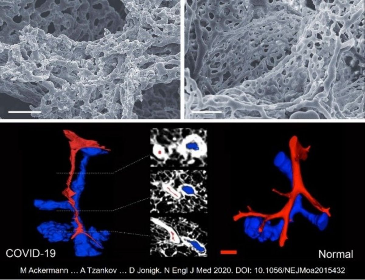
It was the collected material from Basel to allow uncovering of rater unique and distinctive vascular features consisting of severe endothelial injury, associated with intracellular SARS-CoV-2, wide spread thrombosis, microangiopathy and new vessel growth-predominantly by the mechanism of intussusceptive angiogenesis (Figure 2), allowing to concluding that COVID-19 is an angiocentric clinical syndrome [Ackermann et al].

Figure 2. ligne supérieure : micrographes électroniques à balayage des clichés de corrosion microvasculaire du plexus alvéolaire à paroi mince d'un poumon blessé par COVID-19- (à gauche) et d'un poumon sain (à droite).
Lower row : (Sub)total occlusion of the arteries (red) in COVID-19-lung (left), as compared to uninfected controls (right). (μCT 3D reconstruction)

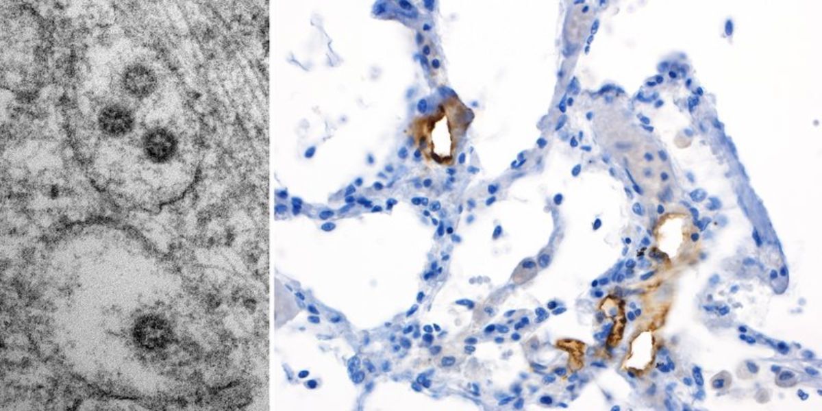
Figure 3 : Images en microscopie électronique de structures virales de taille caractéristique (75-120nm) à l'intérieur des structures intracellulaires membranaires de l'appareil trans-Golgi/du réticulum endoplasmique avec des structures nucléocapsides caractéristiques sur la face interne (~20nm mesurant des points sombres), et des projections environnantes seulement perceptibles. Droite : Staining immunohistochimique d'un échantillon de poumon pour la protéine spike du SRAS-CoV-2 avec le clone 007 de Sino Biological sur un cas autopsié de notre cohorte publiée montrant la présence de virus spikés principalement dans le compartiment endothélial.
Indeed, not only are virus-like particles detectable in endothelial cells, but applying anti-SARS-CoV-2-spike-protein antibodies, one will detect highly preferential presence of virus antigens in the endothelial, but not in the epithelial compartment of the alveolar units (Figure 3), arguing in favor of the above hypothesis.
Sur la base de ce qui précède, la proposition de recherche du groupe de recherche COVID-19 à l'institut a été sélectionnée par le Fast Track Call for Acute Global Health Challenges du Botnar Research Centre for Child Health (BRCCH) et s'est vue attribuer 1,3 million de CHF pour les deux ans et demi à venir. Dans une approche interdisciplinaire avec des chercheurs de pathologie, d'immunologie, de médecine interne, de soins intensifs et de neurologie, des connaissances sur la pathogenèse du COVID-19 sont en train d'être générées dans le but final d'affecter les futures interventions médicales plus adaptées pour cette maladie.
Les principaux thèmes de recherche sont les suivants
- Tropisme tissulaire et cellulaire du SARS-CoV-2 et imagerie in situ de réservoirs de coronavirus
- Analyse immunohistochimique des tissus COVID-19 en ce qui concerne les protéines virales, la composition cellulaire inflammatoire et l'altération des composés de la cascade de l'angiotensine.
- Analyse de l'expression de l'ARN par rapport au SRAS-CoV-2 et de l'expression des gènes de l'angiogenèse, du remodelage tissulaire et de l'immunité/inflammation.
- Imagerie tissulaire à haut paramètre de la pathologie coronavirus (CODEX)
Publications sélectionnées
- Tzankov A, Bhattacharyya S, Kotlo K, Tobacman JK. L'augmentation de la chondroïtine sulfate et la diminution de l'arylsulfatase B peuvent contribuer à la pathophysiologie de l'insuffisance respiratoire COVID-19. Pathobiology. 2021 Nov 17:1-11.
- Yekelchyk M, Madan E, Wilhelm J, et al. Flower lose, a cell fitness marker, predicts COVID-19 prognosis . EMBO Mol Med. 2021 Nov 8;13(11):e13714.
- Nakayama T, Lee IT, Jiang S, et al. Determinants of SARS-CoV-2 entry and replication in airway mucosal tissue and susceptibility in smokers . Cell Rep Med. 2021 oct 19;2(10):100421.
- Hozayen SM, Zychowski D, Benson S, et al. Outpatient and inpatient anticoagulation therapy and the risk for hospital admission and death among COVID-19 patients . EClinicalMedicine. 2021 Nov;41:101139. doi : 10.1016/j.eclinm.2021.101139.
- Menter T, Cueni N, Gebhard EC, et al. Case Report : Co-occurrence of Myocarditis and Thrombotic Microangiopathy Limited to the Heart in a COVID-19 Patient. Front Cardiovasc Med. 2021 Jul 29.
- Obermayer A, Jakob LM, Haslbauer JD, et al. Neutrophil Extracellular Traps in Fatal COVID-19-Associated Lung Injury. Dis Markers. 2021 juil. 30.
- Sahanic S, Löffler-Ragg J, Tymoszuk P, et al. The Role of Innate Immunity and Bioactive Lipid Mediators in COVID-19 and Influenza . Front Physiol. 2021 Jul 22;12:688946.
- Menter T, Tzankov A, Bruder E. SARS-CoV-2/COVID-19-Auswirkungen auf den Placenta [Impact du SARS-CoV-2/COVID-19 sur le placenta]. Pathologe. 2021 Jun 11:1-7.
- Wu CT, Lidsky PV, Xiao Y, et al. SARS-CoV-2 infecte les cellules β pancréatiques humaines et provoque un dysfonctionnement des cellules β. Cell Metab. 2021 Aug 3;33(8):1565-1576.
- Boor P, Eichhorn P, Hartmann A, et al. Aspects pratiques des autopsies COVID-19 [Practical aspects of COVID-19 autopsies]. Pathologe. 2021 Mar;42(2):197-207.
- Bösmüller H, Matter M, Fend F, et al. The pulmonary pathology of COVID-19. Virchows Arch. 2021 Feb 19:1-14.
- Haslbauer JD, Matter MS, Stalder AK, et al. Histomorphological patterns of regional lymph nodes in COVID-19 lungs [Schéma de réaction des ganglions lymphatiques locorégionaux dans la zone de drainage des poumons COVID-19]. Pathologe. 2021 Mar;42(2):188-196.
- Zellweger NM, Huber J, Tsakiris DA, et al. Haemophagocytic lymphohistiocytosis and liver failure-induced massive hyperferritinaemia in a male COVID-19 patient. Swiss Med Wkly. 2021 Jan 15;151:w20420.
- Haslbauer JD, Perrina V, Matter M, et al. Retrospective Post-mortem SARS-CoV-2 RT-PCR of Autopsies with COVID-19-Suggestive Pathology Supports the Absence of Lethal Community Spread in Basel, Switzerland, before February 2020. Pathobiology. 2021;88(1):95-105. doi : 10.1159/000512563.
- Menter T, Tzankov A. Les investigations des pathologistes comme clé pour comprendre la maladie à coronavirus en 2019. Pathobiology. 2021;88(1):11-14.
- Reinhold A, Tzankov A, Matter M, et al. Ocular pathology and occasionally detectable intraocular SARS-CoV-2 RNA in five fatal COVID-19 cases. Ophthalmic Res. 2021 Jan 20.
- Glatzel M, Hagel C, Matschke J, et al. Neuropathologie associée à l'infection par le SRAS-CoV-2. Lancet. 2021 Jan 23;397(10271):276.
- Cribiù FM, Erra R, Pugni L, et al. Severe SARS-CoV-2 placenta infection can impact neonatal outcome in the absence of vertical transmission. J Clin Invest. 2021 Jan 26:145427.
- Fuchs V, Kutza M, Wischnewski S, et al. Presence of SARS-CoV-2 Transcripts in the Choroid Plexus of MS and Non-MS Patients With COVID-19. Neurol Neuroimmunol Neuroinflamm. 2021 Jan 27;8(2):e957.
- Lee IT, Nakayama T, Wu CT, et al. ACE2 se localise au niveau des cils respiratoires et n'est pas augmenté par les inhibiteurs de l'ECA ou les ARA. Nature Communications. 2020 Oct 28;11(1):5453.
- Nienhold R, Ciani Y, Koelzer VH, et al. Two disctinct immunopathological profiles in autopsy lungs of COVID-19. Nature Communications. 2020 Oct 8;11(1):5086.
- Frank S. Catch me if you can : SARS-CoV-2 detection in brains of deceased patients with COVID-19. Lancet Neurol. 2020 Oct 5;19(11):883-4.
- Hopfer H, Herzig MC, Gosert R, et al. Hunting coronavirus by transmission electron microscopy - a guide to SARS-CoV-2-associated ultrastructural pathology in COVID-19 tissues. Histopathology. 2020 Sep 27:10.1111/his.14264.
- Menter T, Mertz KD, Jiang S, et al. Placental Pathology Findings during and after SARS-CoV-2 Infection : Features of Villitis and Malperfusion. Pathobiology. 2020 Sep 18:1-9.
- Ackermann M, Verleden SE, Kuehnel M, et al. Pulmonary vascular endothelialitis, thrombosis, and angiogenesis in Covid-19. The New England Journal of Medicine. 2020 383:120-128.
- Deigendesch N, Sironi L, Kutza M, et al. Correlates of critical illness-related encephalopathy predominate postmortem COVID-19 neuropathology . Acta Neuropathologica. 2020 Aug 26;1-4.
- Eckermann M, Frohn J, Reichardt M, et al. 3D virtual pathohistology of lung tissue from Covid-19 patients based on phase contrast X-ray tomography . eLife. 2020 Aug 20;9:e60408. doi : 10.7554/eLife.60408.
- Jamiolkowski D, Mühleisen B, Müller S, et al. SARS-CoV-2 PCR testing of skin for COVID-19 diagnostics : a case report ; The Lancet. 2020 396(10251):598-599
- Löffler KU, Reinhold A, Herwig-Carl MC, et al. Ocular post-mortem findings in patients having died from COVID-19. Ophthalmologe. 2020 117:648-651
- Menter T, Haslbauer JD, Nienhold R, et al. Post-mortem examination of COVID19 patients reveals diffuse alveolar damage with severe capillary congestion and variegated findings of lung and other organs suggesting vascular dysfunction. Histopathology. 2020 77:198-209
- Mohanty SK, Satapathy A, Naidu MM, et al. Severe Acute Respiratory Syndrome Coronavirus-2 (SARS-CoV-2) and Coronavirus Disease 19 (COVID-19) - anatomic pathology perspective on current knowledge ; Diagnostic Pathology. 2020 15:103.
- Tzankov A, Jonigk D. Unlocking the lockdown of science and demystifying COVID-19 : how autopsies contribute to our understanding of a deadly pandemic. Virchows Archiv. 2020 Sep;477(3):331-333.
Récompenses et subventions
Financement :
Collaborations
- Institute of Pathology, Cantonal Hospital Baselland, Liestal, Switzerland
- Unité de soins intensifs et division de médecine interne, Hôpital universitaire de Bâle, Université de Bâle, Bâle
- Institute of Pathology, Hannover Medical School, et le German Center for Lung Research, Biomedical Research in Endstage and Obstructive Lung Disease Hannover, Germany
- Départements de neurochirurgie et de radiologie, Hôpital universitaire de Bâle, Université de Bâle, Bâle, Suisse
- Nolan Lab, Université de Stanford, Stanford, CA, USA
