Urological Tumor Center
Urologists, radiation oncologists, oncologists and representatives of various highly specialized disciplines work together at the Urological Tumor Center to provide patients and referring physicians with uncomplicated access to highly qualified diagnostics and treatment of tumors of the prostate, bladder and kidneys.
We offer rapid and comprehensive diagnostics with subsequent discussion of the findings in our interdisciplinary tumor meeting and with those affected. This results in a broad-based, personalized treatment recommendation for our patients.
The range of services offered by the Urological Tumour Centre includes not only state-of-the-art diagnostics and robotic surgery, highly specialized radiation modalities and innovative drug therapies, but also comprehensive personal advice for patients. The Tumor Center has a joint uro-oncology consultation hour for urology and radiation oncology , in which specialists from these disciplines discuss the findings and treatment plan with the patient. This service is supplemented by comprehensive psychological counseling and support in the event of emotional stress caused by the tumor disease.
Our active participation in national and international clinical studies expands the treatment options for our patients. We are part of the Swiss Group for Clinical Cancer Research (SAKK) and can make innovative therapies, from the latest cancer drugs to immunotherapies, available to cancer patients at the Tumor Center at an early stage.
Kidney cancer
Changes to the kidneys increase with age. Tumors are growths that displace or penetrate the tissue of the affected organ. Tumors can be malignant or benign. The majority of kidney tumors are benign cysts with no pathological value. In this case, treatment is not necessary if there are no symptoms. Occasionally, however, malignant kidney tumors (cancer) develop, which are often found as an incidental finding in modern imaging such as computer tomography.
Since September 2020, the urological tumor center has also been certified as a kidney cancer center by the German Cancer Society (DKG), which has given the urological tumor center the status of a uro-oncological tumor center. DKG-certified centers must prove annually that they meet the technical requirements for the treatment of a tumor disease and also have an established quality management system. By being certified, we guarantee our kidney cancer patients that they will receive treatment that meets high quality standards at every stage of their disease.
Diagnostics
If we suspect that you have kidney cancer, state-of-the-art imaging is used to determine the size and extent of the cancer in three dimensions. We can then print the cancerous kidney as a 3D model to help plan the operation. In selected cases, we also perform a biopsy to examine the tissue sample under a microscope before any surgery.
Therapy
The treatment of kidney cancer requires the cooperation of various specialists. As part of our certified tumor center, every patient with a newly discovered, locally or systemically advanced kidney cancer is discussed on an interdisciplinary basis in order to offer you an individual and optimally adapted therapy.
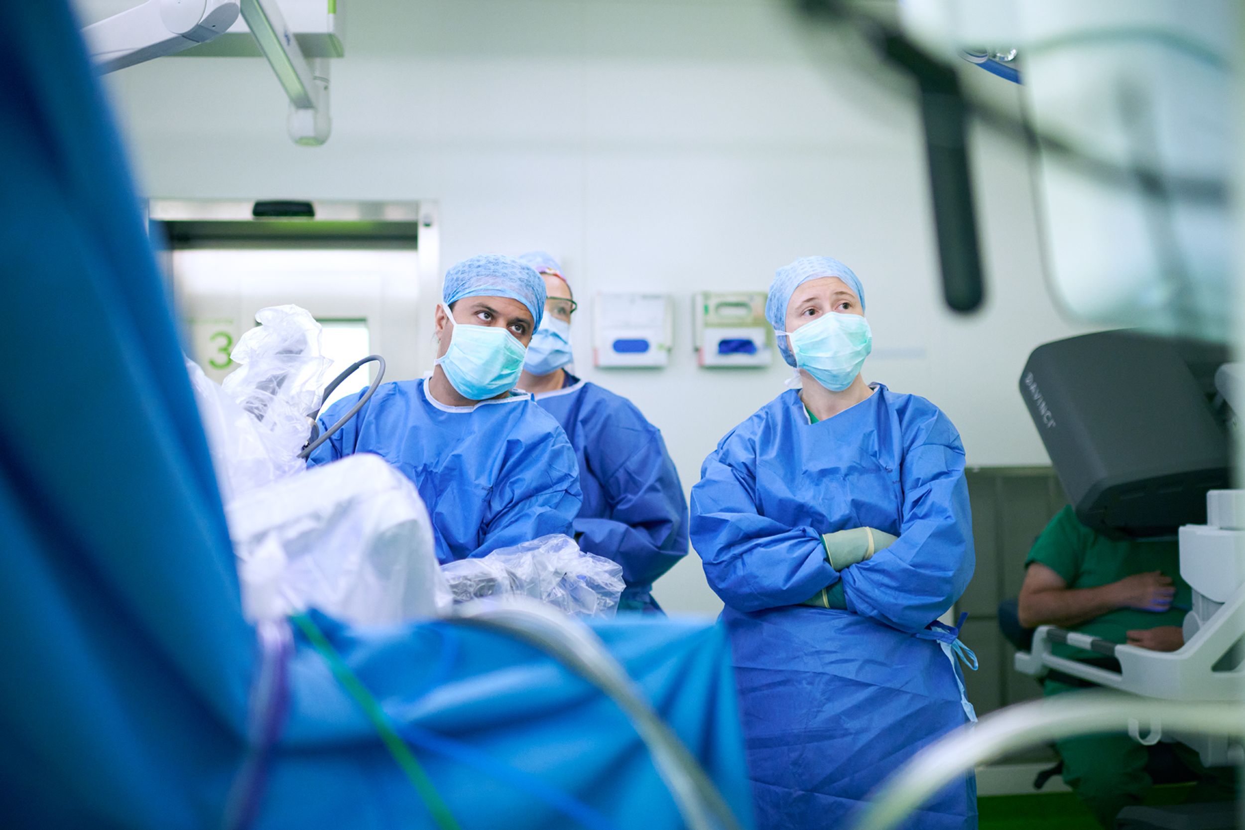
The minimally invasive surgical removal of cancer confined to the kidney is currently the most frequently used and best proven method. Our focus is on minimally invasive, robot-assisted surgery using the Da Vinci® system. Here, the surgeon performs the procedure using microsurgical instruments and a camera, which is held by the robot, through small abdominal incisions. Our experience shows that patients have less pain after this operation, are mobile more quickly and can go home sooner compared to patients who undergo open surgery via a large abdominal incision.
Aftercare
We offer every patient with kidney cancer a tumor follow-up tailored to the patient in order to detect kidney cancer recurrences and possible surgery-related consequences at an early stage and treat them accordingly.
Urinary bladder cancer
Bladder cancer is the fifth most common cancer in humans in Europe. Smokers and men have a significantly higher risk of developing bladder cancer. But exposure to certain chemicals is also a risk factor for the development of bladder cancer. Patients with bladder cancer often report having suffered from bloody urine as the first sign of the disease. However, an increasing urge to urinate without the classic symptoms of bladder inflammation, such as a burning sensation when urinating, can also occur as part of bladder cancer.
Find out more about our expertise in bladder cancer here and gain an insight into the specialist areas of our specialists
Prostate cancer
Prostate cancer is the most common cancer in men.
Precaution
The earlier prostate cancer is detected, the greater the chances of recovery. For this reason, the Swiss Society of Urology recommends prostate cancer screening from the age of 45 if there is a family history of prostate cancer (father or brother with prostate cancer) and from the age of 50 if there is no family history. This screening consists of a palpation of the prostate via the rectum and a blood test (prostate-specific antigen, PSA for short).
Diagnostics
If the screening examination reveals any abnormalities, a tomography scan (magnetic resonance imaging, or MRI for short) of the pelvis is performed to assess the prostate more precisely. If prostate cancer is suspected, tissue samples are taken from the prostate via the rectum under local anesthesia and examined under a microscope. Fusion biopsy, based on imaging using magnetic resonance imaging and transrectal ultrasound (MRT-TRUS), has established itself as a particularly precise method. Suspicious areas of the prostate are visualized using magnetic resonance imaging (MRI) of the pelvis. The magnetic resonance imaging images are superimposed on the ultrasound images of the prostate during the biopsy. This allows the suspicious areas to be precisely targeted for tissue sampling. This method is used as standard at our clinic. With MRI-TRUS fusion biopsy, prostate cancer is diagnosed earlier, more precisely and with fewer tissue samples taken. This means that patients can be given optimal treatment at an early stage and the risk of side effects due to repeated tissue sampling can be reduced.
Therapy
The treatment of prostate cancer requires the cooperation of various specialists. As part of our certified tumor center, every patient with newly discovered prostate cancer is discussed on an interdisciplinary basis in order to be able to offer an individual and optimally adapted therapy. The treatment options available to you will be discussed with you in detail.
Active surveillance
In the case of low-risk prostate cancer that does not exceed the prostate capsule, surgery or radiotherapy can sometimes be dispensed with. Instead, the strategy of active surveillance is used. Prostate cancer is monitored by means of regular check-ups (palpation of the prostate, blood tests and prostate biopsies). This allows changes in the cancer to be detected at an early stage and active therapy to be initiated if the disease progresses. Active monitoring can prevent the side effects of radiotherapy or medication as well as the possible complications of surgery.
Surgical removal of the prostate
Complete surgical removal of the prostate (radical prostatectomy) is recommended for organ-confined cancer growth. This option is currently the most frequently used and very proven method. Our focus is on minimally invasive, robot-assisted surgery using the Da Vinci® system. Here, the surgeon performs the procedure using microsurgical instruments and a camera held by the robot through small abdominal incisions. Our experience shows that patients have less pain after this operation, are mobile more quickly and can go home sooner compared to patients who are operated on openly through a large abdominal incision.
Radiotherapy
Radiotherapy can damage the cancer cells to such an extent that they die. The surrounding healthy organs, such as the small intestine, bladder and genital organs, are spared as much as possible through targeted radiation.
Hormone and chemotherapy
The sex hormone testosterone influences the growth of prostate cells and therefore, under certain circumstances, the growth of prostate cancer. The influence of testosterone on the growth of hormone-dependent prostate cancer is eliminated with anti-hormone therapy. This can be achieved with surgery (subcapsular orchiectomy) or drug treatment.
Chemotherapy can be used for prostate cancer if the anti-hormone therapy is not (or no longer) effective. Chemotherapy is a treatment with cell-damaging or growth-inhibiting drugs. It ensures that fast-growing cancer cells no longer divide and the cancer can therefore no longer multiply. However, chemotherapy also damages healthy, fast-growing cells (e.g. bone marrow cells, hair follicle cells or cells of the mucous membranes in the mouth, stomach or intestines).
Aftercare
After prostate cancer treatment, we offer all patients individual tumor follow-up care and, if available, advice on urinary incontinence and erectile dysfunction.
Testicular cancer
Testicular cancer is one of the most common malignant diseases in younger men. A cure can be achieved in over 95% of cases. The first sign patients often feel is a painless hardening in the area of the testicle. It is important to intervene early in the course of the disease to prevent cancer cells from spreading to the rest of the body.
Diagnostics
If testicular cancer is suspected, the testicles are palpated, an ultrasound examination of the testicles is carried out and special parameters are determined in the blood, which can be elevated in testicular cancer. If these examinations reveal any abnormalities, the testicle must be removed from the scrotum for further diagnosis in the operating room.
Therapy
Treatment planning takes place in a special interdisciplinary conference with colleagues from oncology and radiology. The therapy depends on the type and stage of the testicular cancer, the accompanying illnesses and the patient's personal wishes. As a first step, the affected testicle is usually removed via an incision in the groin. This is sometimes followed by chemotherapy or radiotherapy. A complete cure is often possible in this way, even in advanced stages of cancer.
Aftercare
Once treatment has been completed, regular follow-up checks are necessary in order to counteract a possible relapse at an early stage. In this regard, we will discuss and draw up an individual tumor follow-up plan for you.
Penile cancer
Penile cancer is a rare disease that mostly affects older men. However, every fifth person affected is younger than 60. Known risk factors are infections with the human papillomavirus (HPV) and chronic inflammation of the penis. HPV infection can occur through sexual contact with an infected partner. There are various HPV subtypes, most of which are harmless. However, some are associated with an increased risk of developing penile cancer. Chronic inflammation of the penis can be caused by poor hygiene, foreskin constriction or infections.
Diagnostics
Changes to the testicles and penis are often accompanied by a great sense of shame. It is not uncommon for penile cancer to only be seen by a doctor at a very advanced stage of the disease. At the beginning there is usually a painless reddish or white spot, which is sometimes raised and can quickly increase in size. Sometimes the diagnosis can be made more difficult by a narrowing of the foreskin covering the cancer.
If penile cancer is suspected, a small tissue sample of the findings is usually taken under local anesthesia to confirm the diagnosis. In addition, cross-sectional imaging is carried out to determine the exact extent of the cancer and to look for any offshoots in the body.
Therapy
The therapy depends on the stage of the cancer. Possible treatment options are discussed on an interdisciplinary basis at our tumor board and then discussed with you in detail. The cornerstone of penile cancer therapy is surgery. If possible, the aim is to preserve the penis.
Aftercare
Once treatment has been completed, regular follow-up checks are necessary in order to counteract a possible relapse at an early stage. We will draw up an individualized cancer aftercare plan for you.
Psycho-oncological care
A cancer diagnosis can have a serious impact on your life. The emotional stress during treatment often has an impact on your health-related quality of life. We therefore offer professional support from psychologists specializing in cancer.

Care
Our nursing care at the Urological Tumor Center includes the following
Main focus ward:
- Nursing care and support before and after surgery
- Training on prophylactic measures to avoid complications (e.g. thrombosis prophylaxis or pneumonia prophylaxis)
- Care of urinary drainage systems and wound care. If necessary, in cooperation with the stoma consultation.
- Visits by nursing staff responsible for the ward to discuss and evaluate needs.
- Close interdisciplinary cooperation with other nursing services (APN continence promotion, oncology nursing program management)
Continence promotion (outpatient / inpatient):
Advice for patients, relatives and carers on dealing with urinary diversion systems (transurethral and suprapubic urinary catheters).
- Advice on the problem of incontinence
- Clarification of individually adapted aids such as absorbent incontinence products, urinal condoms or urine bags
- Training of patients and relatives, for example in self-catheterization or in the use of urinary drainage systems (urinary catheters)
- Nursing support and advice after urological procedures such as neobladder, prostatectomy, etc.
- APN continence support (outpatient / inpatient)
Management


Center coordination

PD Dr. Jan Ebbing
Leitender Arzt
Urologie
Teamleiter Robotische Chirurgie der Harnblase
Management body

Prof. Daniel Boll
Stv. Chefarzt Radiologie und Nuklearmedizin
Leitung abdominelle und onkologische Diagnostik, med. Dienstleistung
Tel. +41 61 328 63 84

Prof. Lukas Bubendorf
Leitender Arzt und Fachbereichsleiter Zytopathologie
Pathologie
Tel. +41 61 328 78 51


Prof. Dr. Frank Stenner
Stv. Chefarzt
Innere Medizin (D,CH), Hämatologie (D,CH), Onkologie (CH)
Administration


