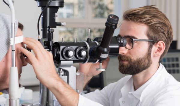
Offer
In addition to our polyclinic for basic care with a 24-hour emergency service, we offer all special consultation hours in ophthalmology. If an operation is necessary, you can rely on our experienced specialists and state-of-the-art technology. In addition to the outpatient area, our clinic also has a ward to ensure you receive the best possible care.
Eye emergency

Was muss ich beachten, wenn ich auf den Augennotfall komme?
- Der Augennotfall ist rund um die Uhr an 7 Tagen pro Woche das ganze Jahr geöffnet.
- Vor allem am Wochenende (Samstagvormittag und Sonntag) müssen Sie mit längeren Wartezeiten rechnen.
- Es werden keine Termine für den Augennotfall vergeben.
Kontakt Augennotfall
Der Augennotfall steht rund um die Uhr an der Mittleren Strasse 91 in Basel zur Verfügung.
Bei Fragen oder im Zweifel, insbesondere auch nachts, können Sie uns anrufen und sich beraten lassen.
Tel. +41 61 265 86 86
Special consultation hours
Registration for consultation hours
Monday through Friday
8.00-12.00 and 13.00-17.00
Augenlid- und Tränenwegssprechstunde
Gesunde und funktionierende Ober- und Unterlider sowie die normale Benetzung der Augen sind für gutes Sehen (und Aussehen) unerlässlich.
Die Schwerpunkte der Augenlid- und Tränenwegssprechstunde sind:
- Kosmetische Lidoperationen (Schlupf-, Hängelider)
- Diagnostik, Behandlung und Entfernung von gut- und bösartigen Tumoren der Augenlider und Orbita
- Rekonstruktion der Augenlider oder Teile des Lids nach Unfällen oder Tumoren
- Wiederherstellung der korrekten Lidstellung und -funktion bei ein- oder ausgedrehten Lidern
- Behandlung chronisch tränender Augen mittels ambulanter Laserchirurgie oder chirurgischen Methoden

Prof. Claudia Auw-Hädrich
Kaderärztin

Dr. Daniela Suter-Starosta
Oberärztin

Dr. Angelika Jähnig
Oberärztin
Endokrine Orbitopathie
Augenbeschwerden durch eine Entzündung der Weichteile der Augenhöhle, die sogenannte Endokrine Orbitopathie, finden sich bei etwa 50% der Patientinnen und Patienten mit einer Autoimmunerkrankung der Schilddrüse (Typ Basedow oder Typ Hashimoto).
Das sehr facettenreiche Krankheitsbild bedeutet für die Betroffenen oft eine massive Einschränkung der Lebensqualität, bedingt durch mögliche Sehstörungen/Doppelbilder, schmerzhafte Entzündungen und äusserliche Veränderungen des Erscheinungsbildes («grosse Augen»).
Ausschlaggebend für eine effektive Behandlung ist die Erkennung der Erkrankung im Frühstadium, das rechtzeitige Einleiten einer eventuell erforderlichen anti-entzündlichen Therapie und die enge Zusammenarbeit mit Kollegen angrenzender Fachgebiete wie Endokrinologie, Oto-Rhino-Laryngologie (HNO) und Mund-Kiefer-Gesichtschirurgie.
Dr. Daniela Suter-Starosta
Oberärztin
Erbliche Netzhauterkrankungen (IRDs, inherited retinal diseases)
In der Sprechstunde für erbliche Netzhauterkrankungen (IRDs, inherited retinal diseases) untersuchen und behandeln wir Patientinnen und Patienten mit erblichen Netzhauterkrankungen, toxischen/Medikamenten-bedingten Netzhauterkrankungen sowie seltenen Autoimmunerkrankungen der Netzhaut.
Zur Diagnostik ihrer Netzhaut-Struktur und -Funktion setzten wir in dieser Sprechstunde ein Spektrum an nicht-invasiven Untersuchungsmethoden ein. Dies umfasst unter anderem hochauflösende optische Kohärenztomographie («3D-Bildgebung des Augenhintergrunds»), Fundusautofluoreszenz («metabolische Bildgebung»), Gesichtsfeldmessungen sowie Elektroretinographie («Ableitung der im Auge erzeugten elektrischen Spannung»).
Für eine optimale Versorgung unserer Patientinnen und Patienten arbeiten wir eng mit der Genetik Forschungsgruppen von Professor C. Rivolta zusammen. Dies ermöglicht, dass Patientinnen und Patienten von molekulargenetischen Untersuchungen auf dem neuesten Stand der Wissenschaft profitieren.
Wir bieten die Behandlung mit zugelassenen Therapien (Gentherapie Voretigene neparvovec, Transkorneale Elektrostimulation) an, und vermitteln humangenetische Beratung, sowie Low-Vision und soziale Beratung. Einmal monatlich findet zudem in der Augenklinik Basel eine Beratungssprechstunde der Retina Suisse (https://retina.ch/) statt.
An der Augenklinik Basel wird Therapieforschung für erbliche Netzhauterkrankungen durchgeführt. Hierdurch können Patientinnen und Patienten von neusten Therapien vor Marktzulassung profitieren. Sollte die Möglichkeit der Teilnahme an einer Therapie-Studie bestehen, würden wir Sie hierzu, sowie den möglichen Nutzen und Risiken informieren.
Dr. Virginie Peter
Assistenzärztin
Glaukom
In der Glaukom Sprechstunde, mit langer Tradition in der Augenklinik des Universitätsspitals Basel, wird ein holistisches Prinzip der Betreuung von Patientinnen und Patienten mit Glaukom aller Arten angewendet. Wir bemühen uns aus dem Begriff «Patientinnen und Patienten, und nicht nur die Erkrankung, behandeln» nicht nur eine hohle Phrase zu machen. Patient*innen mit Offenwinkelglaukom, Pseudoexfoliations- und Pigmentdispersionsglaukom, Normaldruckglaukom, Engwinkelglaukom, Neovaskularisationsglaukom und mit Myopie assoziiertem Glaukom erhalten bei uns sowohl diagnostisch als auch therapeutisch – medikamentös, Laser, chirurgisch – Verfahren auf universitärem Niveau und im Einklang mit den aktuellen Erkenntnissen kommuniziert an Kongressen, aus der Forschung und aus internationalen Studien.
«Das» Glaukom gibt es nicht, und das wissen wir. Ein Glaukom ist individuell, und bei jeder Person ist die Zusammensetzung der Risikofaktoren, die zu einem beschleunigten Verlust von wertvollen retinalen Ganglienzellen und somit Axonen des Sehnervs führen, anders. Wir besitzen sowohl Erfahrung als auch Expertise auf höchstem Niveau, und ebenfalls die modernste - auch bildgebende - Technologie, ein strukturiertes Risikoprofil bei einzelnem Betroffenen personalisiert zu erstellen. Basiert darauf, wir setzen den therapeutischen Fokus – den Augendruck akut und/oder langfristig, pharmakologisch, mit Laser oder chirurgisch zu senken; optimale Bedingungen für die Sehnervdurchblutung zu erreichen; retinale Ganglienzellen zu schützen.
Der Gipfel dieser Strategie, was seit mehr als 30 Jahren bei besonders komplexen klinischen Bilder eines Glaukoms in Basel praktiziert wird, ist eine Abklärung des Risikoprofils unter stationären Bedingungen, wo die grosse Mehrheit bekannter und möglichen Risikofaktoren aufs Mal, fokussiert und, wichtig, parallel erfasst und analysiert wird.
Die Glaukom Sprechstunde arbeitet eng zusammen mit niedergelassenen Kolleginnen und Kollegen aus der Region, aus der ganzen Schweiz und auch aus Ausland, und diese Kooperation zu dem Wohle und zu der langfristig bestmöglichen Betreuung unserer Glaukompatient*innen ist unser höchstes Gut.
Prof. Konstantin Gugleta
Stv. Klinischer Chefarzt
Facharzt Ophthalmologie und Ophthalmochirurgie
Hornhaut und Katarakt
Die Sprechstunde Hornhaut und Katarakt beinhaltet die Diagnostik sowie die medikamentöse und chirurgische Behandlung der Erkrankungen der brechenden Medien des vorderen Augenabschnittes:
- der Hornhaut (Cornea)
- der Linse
Grauer Star-Chirurgie:
Trübungen der Linse (Grauer Star/Katarakt) werden mit modernen Operationsmethoden, u.a. mittels Femtosekundenlaser, durch Einsetzen einer Kunstlinse behandelt. Die bei uns implantierten monofokalen Kunstlinsen sind standardmässig asphärisch, haben einen Blaufilter und sind ohne Aufpreis. Zudem bieten wir gegen Aufpreis eine Vielfalt an Premium-Intraokularlinsen wie z.B. multifokale Linsen zur Brillenfreiheit und torische Kunstlinsen zur Astigmatismuskorrektur sowie Speziallinsen bei besonderen Vorderaugenabschnittssituationen (z.B. irreguläre Hornhaut, Aphakie, Aniridie). Die Implantation der Premium Linsen erfolgt typischerweise mittels Femtosekundenlaser unter digitaler Mikroskopie mit computerassistierter Navigation zur maximalen Präzision, Sicherheit und Effizienz.
Hornhautchirurgie:
Bei Hornhautquellung aufgrund verschiedener Hornhautendotheldystrophien wie z.B. Fuchs’scher Endotheldystrophie, oder bei St. n. Verätzung, komplizierter intraokulärer Operation, wird eine schonende, minimal invasive Hornhautschichttransplantation durchgeführt (Descemet Membrane Endothelial Keratoplasty - DMEK), welche durch das Ersetzen der hinteren Hornhautschicht (Hornhautendothel) eine schnelle visuelle Rehabilitation gewährleistet.
Bei Hornhauterkrankungen mit Aussparung des Hornhautendothels wie z.B. bei fortgeschrittenem Keratokonus, wird eine tiefe anteriore lamelläre Keratoplastik (DALK) durchgeführt, bei welcher nur der anteriore Hornhautteil ausgetauscht wird.
Wenn die volle Hornhautdicke vom Epithel bis zum Endothel hin betroffen ist, wie z.B. bei tiefgreifenden Hornhautnarben bei St.n. Hornhautinfektionen, besteht die Möglichkeit einer perforierenden Keratoplastik (PKP).
Bei allen Arten der Hornhauttransplantation und insbesondere bei den lamellären Transplantationstechniken werden wir durch Femtosekundenlaser-Einsatz und digitaler Mikroskopie mit intraoperativer OCT-Aufnahme unterstütz, welche eine maximale Präzision und Sicherheit, insbesondere bei komplexen Fällen, gewährleisten.
Schliesslich, werden komplexe Vorderaugenabschittsdefekte mittels hochspezialisierter Rekonstruktionen repariert. Diese umfassen Interventionen auf der Ebene der Hornhaut (Keratektomie, Amniomembrandeckung, Keratoplastik, ect.), der Iris (Iridoplastiken verschiedener Art, künstliche Iris-Implantation) und der Linse (Implantation von Speziallinsen).
Kataraktoperation in der Augenklinik des Universitätsspital Basel (Video)

Prof. Zisis Gkatzioufas
Leitender Arzt
Facharzt Ophthalmologie und Ophthalmochirurgie
Dr. Oussama Habra
Oberarzt
Dr. Eleftherios Chatzimichail
Oberarzt
Klinische Pathologie / Tumorsprechstunde
Die Sprechstunde richtet sich an Patient*innen mit gutartigen und bösartigen Tumoren des Augapfels, der Augenlider, der Bindehaut, des Tränenwegsystems und der Augenhöhle.
Tumoren stellen oft eine komplexe Erkrankung dar. Eine sorgfältige Diagnosestellung ist unabdingbar. Dazu stehen uns modernste Untersuchungsmethoden zur Verfügung, so bieten wir unter anderem den klassischen Ultraschall auch eine Duplexsonographie und Ultraschallbiomikroskopie an. Je nach Tumor ist eine alleinige Behandlung in der Augenklinik möglich oder eine interdisziplinäre Zusammenarbeit von hoher Bedeutung mit Besprechungen an Tumorboards im Universitätsspital Basel im Speziellen Kopf-, Hals-, Augentumoren. Wir pflegen eine Kooperation mit dem Paul-Scherrer-Institut in Villigen. Uns ist es ein grosses Anliegen modernste und eine individuell angepasste Behandlung nach neusten wissenschaftlichen Erkenntnissen anzubieten.
Prof. Dr. Peter Meyer
Leitender Arzt
Facharzt Ophthalmologie und Ophthalmochirurgie
Prof. Claudia Auw-Hädrich
Kaderärztin
Dr. Elisabeth Graeff
Oberärztin
Dr. Alexandra Steinemann-Inauen
Oberärztin
Kontaktlinsen
Die Zusammenarbeit zwischen Kontaktlinsenoptiker*innen und der Augenklinik ist von grosser Bedeutung. Hier passen wir die verschiedenen Kontaktlinsentypen fachgerecht an.
In der Kontaktlinsen-Sprechstunde bieten wir unseren Patientinnen und Patienten
- Standardanpassungen
- die Korrektur komplizierter Sehfehler und unregelmässiger Hornhautoberflächen mit speziell angefertigten Kontaktlinsen
- therapeutische Kontaktlinsen
- individuelle Beratung
Nebst ausgesuchtem Kontaktlinsenzubehör steht zudem ein Sortiment an Pflegemitteln zu Verfügung.
Ralf Beuschel
Kontaktlinsenspezialist
Medical Retina
In der Medical Retina werden sämtliche Erkrankungen der Netzhaut diagnostiziert und behandelt, bei denen primär eine medikamentöse oder Laser-chirurgische Therapie im Vordergrund steht. Bei speziellen Befunden wie erblichen Netzerkrankungen oder chirurgischen Fragestellungen verweisen wir Sie an unsere hierzu bestehenden Spezialsprechstunden weiter.
Insbesondere Erkrankungen wie die altersabhängige Makuladegeneration (AMD), diabetische Retinopathie, arterielle und venöse Gefässverschlüsse, sowie Netzhaut-Veränderungen bei Kurzsichtigkeit (myope Retinopathie) stehen in der Medical Retina im Vordergrund.
Expertise auf höchstem Niveau: Unsere spezialisierten Fachärzte sind führend in der Forschung und Behandlung von Netzhauterkrankungen. Wir nehmen an internationalen Behandlungsstudien teil und sind auf Kongressen aktiv vertreten. Sie profitieren von der Expertise und dem Wissen, das nur an einem Universitätsspital verfügbar ist.
Modernste Technologie: Wir verwenden die neueste Diagnostik- und Behandlungstechnologie, um präzise Diagnosen zu stellen und effektive Therapien anzubieten. Unsere Diagnostikabteilung verfügt über die neueste Generation von Bildgebungsgeräten. Hiermit können wir ultrahochauflösende Bilder erzeugen und Erkrankungen in frühesten Stadien erkennen. Unser Ziel ist es, Ihre Sehkraft bestmöglich zu schützen und zu verbessern.
Individuelle Betreuung: Jede Patientin und jeder Patient erhalten eine massgeschneiderte Behandlung, die auf ihre individuellen Bedürfnisse abgestimmt ist. Wir nehmen uns Zeit für Sie und beantworten all Ihre Fragen ausführlich.
Umfassendes Leistungsspektrum: Von der Diagnostik über die Therapie bis hin zur Nachsorge bieten wir ein umfassendes Spektrum an Dienstleistungen zur Behandlung von Erkrankungen wie die altersabhängige Makuladegeneration (AMD), diabetischer Retinopathie, Gefässverschlüsse und Myopie an. Intravitreale Injektionen (anti-VEGF-Therapie) zur Behandlung der feuchten Form von AMD und anderen Erkrankungen werden mehrmals pro Woche angeboten.

Prof. Nicolas Feltgen
Klinischer Chefarzt
PD Dr. Josep Callizo
Leitender Arzt
Neuroophthalmologie
Die Neuroophthalmologie befasst sich mit Erkrankungen des Nervensystems in Bezug auf die Zusammenarbeit der Augen und die Verarbeitung von Seheindrücken. Diese gehen oft mit Sehstörungen wie z.B. Sehschärfeverlust, Gesichtsfeldausfall und Doppelbildern einher. Ursache sind Erkrankungen des Sehnervs, der Sehbahn, der verarbeitenden Sehzentren im Gehirn oder der Hirnareale, welche die Augenbewegungen steuern. Auch Symptome wie unterschiedlich grosse Pupillen oder ein Augenzittern können auf ein neuroophthalmologisches Problem hinweisen.
Prof. Anja Palmowski-Wolfe
Leitende Ärztin
Fachärztin Ophthalmologie und Ophthalmochirurgie
Netzhaut- und Hinterabschnitt-Sprechstunde
Die Sprechstunde Hinterabschnitt beinhaltet die Diagnostik sowie die medikamentöse und chirurgische Behandlung der Erkrankungen der Netzhaut:
Die Behandlung von Netzhauterkrankungen hat sich in den vergangenen Jahren deutlich verbessert. Die meisten Erkrankungen sind mittlerweile behandelbar.
Die Universitäts-Augenklinik in Basel legt besonderen Wert auf die Behandlung von Netzhauterkrankungen und bietet neben einer hochmodernen Diagnostik auch das gesamte therapeutische konservative und operative Spektrum.
Zahlreiche spezialisierte Untersuchungsmethoden ermöglichen eine präzise Diagnosestellung. Wir bieten unseren Patienten eine umfassende Beratung über die Therapiemöglichkeiten an.
Netzhauterkrankungen, die eine Operation erfordern:
Operationen am Auge sind heutzutage sehr standardisiert und sicher. Gerade an der Netzhaut sind viele Erkrankungen nur mit einer Operation zu behandeln. Diese werden minimal invasiv durchgeführt.
Prof. Nicolas Feltgen
Klinischer Chefarzt
PD Dr. Josep Callizo
Leitender Arzt
Orthoptik und Sehschule
Die Sprechstunde richtet sich an Kinder und Erwachsene mit Problemen der Zusammenarbeit beider Augen. Ein engagiertes Team aus Ärzt*innen und Orthoptist*innen betreut unsere Patientinnen und Patienten, bei komplexen Erkrankungen in enger Zusammenarbeit mit den Kolleginnen und Kollegen des Universitäts-Kinderspital Beider Basel (UKBB) und des USB.
Schielbehandlung (Orthoptik)
Ein Schielen wird auf der Orthoptik auch im Hinblick auf die Ursache und die Sehentwicklung genauestens untersucht, Für jeden Patient wird individuell die beste Behandlung geplant. Falls notwendig wird eine Schieloperation durchgeführt. Diese erfolgt ambulant und bei Kindern in Vollnarkose. Bei geeigneten erwachsenen Patienten kann eine Schieloperation gerne auch in örtlicher Betäubung unter Gabe von Beruhigungsmitteln (Sedation) erfolgen.
Kinderophthalmologie
In dieser Sprechstunde geht es um das rechtzeitige Erkennen und die Behandlung von Augenerkrankungen im Kindesalter. Je nach Diagnose werden unterschiedliche Fachrichtungen hinzugezogen.
Ein grosser Schwerpunkt liegt auf der Abklärung von Sehbeeinträchtigungen. Dazu zählen vererbbare Augenerkrankungen bei Kindern, ebenso wie die im Vergleich häufiger auftretende Schwachsichtigkeit, die durch gezielte (Vorsorge-)Untersuchungen rechtzeitig erkannt, behandelt und im Kindesalter sogar geheilt werden kann. Auch Kopfschmerzerkrankungen können u.a. durch Bestimmung der Brechkraft des Auges, des Gesichtsfelds und des Sehnerven weiter abgeklärt werden. Unser Spektrum beinhaltet ebenfalls Vorsorgeuntersuchungen bei Neu- und Frühgeborenen.
Uns stehen verschiedene Methoden zur Prüfung der Sehschärfe auch bei Babys und Kleinkindern, sowie bei Patienten mit speziellen Bedürfnissen zur Verfügung. Ebenso können wir auch in diesen Patientengruppen bereits ermitteln, wie die Brechkraft des Auges ist, d.h. ob das Tragen einer Brille nötig ist, um eine für die Entwicklung einer guten Sehschärfe notwendige klare Abbildung eines Bildes auf der Netzhaut zu erreichen. Die Untersuchungen an Kindern finden in kindgerechtem Umfeld mit Spiel- und Wickelmöglichkeit statt.
Myopiesprechstunde (Kurzsichtigkeit)
Mit zunehmender Kurzsichtigkeit steigt das Risiko später im Alter Folgeerkrankungen zu erleiden.
Ganz stoppen kann man die Entwicklung zur Kurzsichtigkeit nicht, aber inzwischen stehen uns Möglichkeiten zur Minderung des Fortschreitens zur Verfügung (Spezialbrillen, Spezialkontaktlinsen, Augentropfen). Um dieses optimal umsetzen zu können, und auch mit einer Kurzsichtigkeit einhergehende andere Erkrankungen rechtzeitig zu erkennen, bieten wir eine Spezialsprechstunde zur Myopie.
Botox Sprechstunde
Diese Sprechstunde richtet sich in erster Linie an Patienten mit unwillkürlichen Muskelkrämpfen im Gesicht (Dystonien), zB. unwillkürlichem Lidkrampf (Blepharospasmus), Meige Syndrom oder Hemispasmus facialis.
Botox wird auch zur Behandlung eingesetzt bei:
- gewissen Schielformen
- Oberlidretraktion (zu weit geöffnetem Oberlid),
- Fehlendem Lidschluss mit daraus folgender Hornhautaustrocknung
- Stark tränendem Auge
Mo–Fr 8.00–12.00 / 13.30–16.30 Uhr
Prof. Anja Palmowski-Wolfe
Leitende Ärztin
Fachärztin Ophthalmologie und Ophthalmochirurgie
Dr. Françoise Roulez
Spezialärztin
Dr. Daniela Suter-Starosta
Oberärztin
Dr. Isabel Kloos
Stv. Oberärztin
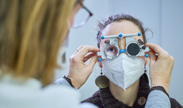
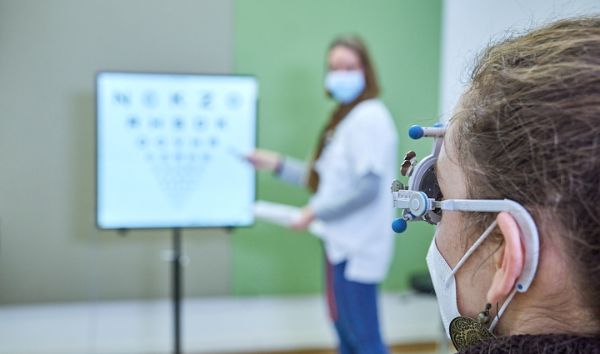
Refraktive Sprechstunde
Die Refraktive Sprechstunde befasst sich mit diagnostischen und therapeutischen Ansätzen, welche als Ziel haben, den Bedarf an optischer Korrektur zu eliminieren und dadurch ein perfektes Sehen ohne Brille oder Kontaktlinse zu ermöglichen.
Refraktive Chirurgie:
Refraktive Fehler wie z.B. Kurzsichtigkeit, Astigmatismus oder Weitsichtigkeit werden mittels Laserbehandlungen (transPRK, Femto-LASIK) bzw. mittels refraktives Linsenaustausches (RLE) korrigiert, damit eine nachhaltige Brillenfreiheit erreicht werden kann. Bei hohen refraktiven Fehlern, bieten wir die Implantation von Implantierbaren Collamer Linsen (ICL). Schliesslich bieten wir bei Keratokonus, ganz elektiv und individuell, eine visuelle Rehabilitation mittels minimal invasiver Methoden, wie z.B. Femtosekundenlaser-assistierter Implantation von kornealen Ringsegmenten (CAIRS, KeraNatural) oder spezieller Laserbehandlungen (customized corneal surface ablation) meistens kombiniert mit kornealem Crosslinking (CXL).
Prof. Zisis Gkatzioufas
Leitender Arzt
Facharzt Ophthalmologie und Ophthalmochirurgie
Dr. Oussama Habra
Oberarzt
Dr. Eleftherios Chatzimichail
Oberarzt
Sicca
Das Sicca-Syndrom ist eine komplexe Erkrankung, die eine gestörte Funktion der Tränen- und Speicheldrüsen zur Folge hat. Zu den häufigsten Beschwerden gehören trockene Augen, Mundtrockenheit, Reizungen der Schleimhäute und eine erhöhte Anfälligkeit für Infektionen. Die Ursachen sind oft vielfältig und reichen von Autoimmunerkrankungen über Umweltfaktoren bis hin zu Nebenwirkungen von Medikamenten. Entscheidend für eine erfolgreiche Behandlung sind eine fundierte Diagnostik und eine individuell angepasste Therapie.
Unser Angebot in der Sicca-Sprechstunde:
Wir bieten eine umfassende Diagnostik, um die Ursache der Beschwerden zu ermitteln und den bestmöglichen Therapieansatz zu finden. Unsere diagnostischen Verfahren umfassen:
- Meibographie (OCULUS Keratograph® und LipiView®) zur Beurteilung der Meibom-Drüsenfunktion und und der Lipidschicht.
- Schirmer-Test zur Messung der Tränenproduktion.
- Tränenfilmanalyse mit Tränenaufrisszeit zur Beurteilung der Stabilität des Tränenfilms.
Auf Basis der diagnostischen Ergebnisse wird die Therapie individuell angepasst. Unsere Therapieoptionen umfassen:
- Breites Angebot und Information über Tränenersatzmitteln, die den unterschiedlichen Bedürfnissen der Patienten gerecht werden.
- Medikamentöse Therapie, wie Ciclosporin zur anti-inflammatorischen Therapie oder Eigenserum-Augentropfen, bei Bedarf zur Unterstützung der Heilung der Augenoberfläche.
- Wärmebehandlung der Lider mittels Blepha-Steam Brille (Thea®), zur Behandlung der weit verbreiteten Meibomdrüsen-Dysfunktion.
- Professionelle Lidreinigung zur Reduzierung von Lidrandentzündungen und Verbesserung der Tränenfilmqualität.
- Punctum Plugs zur Reduzierung des Tränenabflusses und Verlängerung der Tränenfilm-Stabilität.
- Neu: IPL-Lasertherapie (Intense pulsed light, Lumibird®) zur Behandlung der Augenlider bei chronischer Liddrandentzündung und Meibomdrüsendysfunktion.
Zusätzlich führen wir eine strukturierte Anamnese durch, bei der mit Hilfe des adaptierten OSDI-Fragebogens (Ocular Surface Disease Index) spezifische Symptome und Beschwerden erfasst werden. Dies ermöglicht eine detaillierte Einschätzung des Schweregrades der Erkrankung.
Prof. Claudia Auw-Hädrich
Kaderärztin
Dr. Angelika Jähnig
Oberärztin
Uveitis
Das Wort «Uvea» bezeichnet die mittlere vaskularisierte (gut durchblutete) Schicht im Auge; Uvea setzt sich aus der Regenbogenhaut, dem Strahlkörper, und der Aderhaut zusammen. All die drei teilen können entzündet werden, daher das «-itis» Suffix. Jede Entzündung verursacht Rötung, Wärme, Schmerz, Schwellung und Funktionsreduktion, und eine – oder mehrere – von diesen Symptomen führen Patienten zum Augenarzt. Die Aufgabe von Augenärztin in dieser Situation ist zweierlei: da das Auge ein Teil des menschlichen Körpers ist und alle – insbesondere immunologische – Störungen sich im Auge widerspiegeln können, zuerst eine mögliche systemische Erkrankung auszuschliessen, oder ggf. aufzuspüren; und, dem/der Patient/in unmittelbar zu helfen, indem die Beschwerden mittels antientzündlicher Pharmakologie gelindert werden.
Was ist die Rolle der Uveitis-Sprechstunde in der Augenklinik des Universitätsspitals Basel? Diagnostik der Uveitis (anteriore / intermediäre / posteriore; Iridozyklitis, Chorioiditis, retinale Vaskulitis usw.) kann sehr komplex werden, und braucht nicht selten ein multidisziplinäres Verfahren. Wir sind eingebettet im Universitätsspital, und haben somit, ebenfalls ad personam, direkten Zugang zu universitären Ressourcen in Fachgebieten der Inneren Medizin, Rheumatologie, Pneumologie, Immunologie, und Pathologie/Onkologie. Auf der Therapie-Seite, insbesondere bei posteriorer Uveitis (Entzündungen der Aderhaut, Netzhaut und Netzhaut-Gefässen), braucht es eine Modulation des Immunsystems. Diese erreicht man heutzutage einerseits mit intraokular applizierten i.d.R. Steroidhaltigen-Implantaten, und wir haben Zugang zu und Erfahrung mit diesen Medikamenten; andererseits, durch die Anwendung von modernen Biologika - Antikörper mit, in diesem Kontext, speziellen Effekten auf das Immunsystem - im Einklang mit den aktuellen Erkenntnissen kommuniziert an Kongressen, aus der Forschung und aus internationalen Studien.
Eine möglichst beste Betreuung von Uveitis Patienten setzt enge Zusammenarbeit nicht nur mit den anderen Fachgebieten und Hausärzte voraus, sondern auch mit anderen Uveitis-Zentren in der Schweiz, und ebenfalls mit niedergelassenen Augenärzten / innen aus der Region, aus der ganzen Schweiz und aus dem Ausland. Diese Kooperation zu dem Wohle unserer Uveitispatienten ist unser höchstes Gut.
Dr. Oussama Habra
Oberarzt
Dr. Alexandra Steinemann-Inauen
Oberärztin
Dr. Jürg Messerli
Arzt
Prof. Konstantin Gugleta
Stv. Klinischer Chefarzt
Facharzt Ophthalmologie und Ophthalmochirurgie
General consultation hours
Hospital specialist team
Our hospital specialists care for patients across the spectrum of eye diseases. The main care model is long-term personal ophthalmological care; this is particularly important for multimorbid patients who need structured regular ophthalmological check-ups and treatment, but who are unable to receive this in a highly specialized consultation due to the many eye diseases present at the same time. If necessary, because it is well integrated into the eye hospital, the hospital specialist team can directly access the relevant specialized expertise.
Kataraktoperation in der Augenklinik des Universitätsspital Basel
Frequently asked questions and answers
"Eye accident - what should I do?"
A small, superficial injury to the cornea or conjunctiva, for example caused by a fingernail, usually heals itself without any major complications. An immediate visit to an eye emergency is usually not necessary. An ophthalmologist's check-up within one or two days is advisable.
In the following cases, please come to our eye emergency center as soon as possible:
- Foreign body in the eye (e.g. metal splinters)
- Chemical burn with liquid - rinse eyes immediately with water and, if possible, describe the liquid in detail (e.g. photo of packet)
- Severe blow to the eye or injury with a sharp object
- Injuries with branches or organic materials
If you have poor vision in the affected eye after the accident, a prompt medical examination is also recommended.
Please refrain from wearing contact lenses after an eye accident.
"My eyes are red and it bites."
If your eyes are red and burning, it is usually conjunctivitis. It is often caused by viruses and is very contagious, but harmless. Normally, you can wait a few days for spontaneous improvement before contacting the eye clinic.
You can obtain advice from us by telephone. As a first measure, you can buy moisturizing eye drops from the pharmacy to alleviate the symptoms ("artificial tears"). Good hygiene reduces the risk of infecting other people: if possible, avoid touching your eyes and wash and disinfect your hands regularly.
In the following cases, please contact the eye clinic on the same day or the next day:
- You wear contact lenses and suspect conjunctivitis. Please do not wear your contact lenses during this time.
- You have redness on one side of your eye with pain and a deterioration in vision.
- You have conjunctivitis, work in a profession with special hygiene regulations or intensive contact with people and require a doctor's certificate.
- You have recently undergone eye surgery.
- You have pain in or around the eye that cannot be alleviated with moisturizing eye drops.
"I can't see so well anymore."
Has your vision suddenly deteriorated in one eye, for example after waking up? Then please come to the eye emergency immediately. You can call beforehand for an initial clarification and assessment.
In the following cases, you should make an appointment at the eye clinic on the same day or the next day:
Your vision has gradually deteriorated and....
- You see shadows, flashes or black dots.
- You can no longer see everything in your field of vision.
- You suffer from diabetes or an eye disease (e.g. age-related macular degeneration AMD).
- You have recently had eye surgery.
If your eyes are dry for a longer period of time, your vision will also deteriorate. Sicca occurs when the wetting of the surface of the eye does not function properly (due to various causes). These symptoms can be treated in our sicca consultation.
If your vision gradually deteriorates in both eyes, this is not usually an emergency. You can register for a comprehensive assessment during our regular consultation hours.
"There's something floating on my eye." "I see flashes."
Please come to our eye emergency center as soon as possible if your vision has deteriorated in one eye and you see flashes or "floating flies".
If you see many black dots ("sooty rain"), this is an indication of a possible retinal tear. In this case, please contact the eye clinic on the same day or the next day.
If your vision has not changed and you see floating particles in your eye ("flying gnats") without flashes, soot rain or a shadow, a visit to the eye emergency clinic is not normally necessary. You can register for a check-up during our consultation hours. Please note that an examination with a wide pupil must be carried out for further clarification and that you will not be able to drive for three to four hours afterwards (please do not come by car, bicycle or other self-driven vehicle).
"My optician says I should have my eyes checked more closely."
Please contact the eye clinic as soon as possible if your optician has recommended a visit to the ophthalmologist. Older people often have cataracts, which can be easily operated on. It is essential that other causes are ruled out, especially in the case of a one-sided deterioration in vision.
"I need new glasses."
We recommend our glasses consultation for people who wear glasses. You can book an appointment for a consultation online. Please contact your health insurance provider if you have any questions about the reimbursement of new spectacle lenses. After eye surgery or chronic eye diseases, insurance companies usually grant a subsidy.
"I would like to switch to contact lenses."
We recommend our consultation hours for contact lenses. You can register via our online appointment booking system. If you have any questions about the reimbursement of contact lenses, please contact your health insurance company.
"I would like to get a second opinion on my diagnosis."
The eye clinic offers special consultation hours for numerous eye diseases. You can make an appointment for a second medical opinion in the special consultation that deals with your disease. You can call us in advance for advice.
Tel. +41 61 265 86 86
Please bring the documents from your doctor with you to the consultation or have them sent to the eye clinic in advance.
"My eyelid is swollen."
A swollen area on the eyelid (urseli, chalazion, sty) is usually harmless and disappears after a few days. An immediate visit in an eye emergency is not usually necessary. You can register for our consultation hours.
If the swelling and redness increases and extends beyond the eyelid or if you are in severe pain, you should visit our eye emergency department as soon as possible.
"One of my eyes is red, but it doesn't itch or hurt."
If you have not received a blow to the eye and have not noticed any change in your vision, it is probably a harmless hemorrhage into the conjunctiva. ("bruise on the eye"). The redness will disappear within a few days to weeks. A visit to your general practitioner to check for a possible underlying increase in blood pressure may be advisable.