Offer
As a center hospital, the University Hospital Basel offers the entire spectrum of urological and uro-oncological procedures. As a specialization, the entire range of minimally invasive surgical methods such as laparoscopy, retroperitoneoscopy, robotic surgery and laser surgery are offered.
Contact for consultation appointments
Urology registration office
Availability: Mon-Fri, 08.00 - 12.00 / 13.00 - 17.00
Phone +41 61 265 72 80
Fax +41 61 265 76 90
anmeldung.urologie@usb.ch
Contact for emergencies
In emergencies, we are there for you around the clock.
Emergency Center
University Hospital Basel Petersgraben 2
4031 Basel
Phone +41 61 265 25 25
Range of services
Tumor consultation hours
Kidney tumor
Changes to the kidneys increase with age. Tumors are growths that displace or penetrate the tissue of the affected organ. Tumors can be malignant or benign. The majority of kidney tumors are benign cysts with no pathological value. In this case, treatment is not necessary if there are no symptoms. Occasionally, however, malignant kidney tumors (cancer) develop, which are often found as an incidental finding in modern imaging such as computer tomography.
Since September 2020, the urological tumor center has also been certified as a kidney cancer center by the German Cancer Society (DKG), which has given the urological tumor center the status of a uro-oncological tumor center. DKG-certified centers must prove annually that they meet the technical requirements for the treatment of a tumor disease and also have an established quality management system. By being certified, we guarantee our kidney cancer patients that they will receive treatment that meets high quality standards at every stage of their disease.
Diagnostics
If we suspect that you have kidney cancer, we use state-of-the-art imaging to determine the size and extent of the cancer in three dimensions. We can then print the cancerous kidney as a 3D model to help plan the operation. In selected cases, we also perform a biopsy to examine the tissue sample under a microscope before any surgery.
Therapy
The treatment of kidney cancer requires the cooperation of various specialists. As part of our certified tumor center, every patient with newly discovered, locally or systemically advanced kidney cancer is discussed on an interdisciplinary basis in order to offer you an individual and optimally adapted therapy.
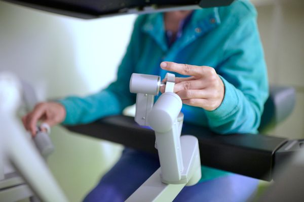
The minimally invasive surgical removal of cancer confined to the kidney is currently the most frequently used and best proven method. Our focus is on minimally invasive, robot-assisted surgery using the Da Vinci® system. Here, the surgeon performs the procedure using microsurgical instruments and a camera, which is held by the robot, through small abdominal incisions. Our experience shows that patients have less pain after this operation, are mobile more quickly and can go home sooner compared to patients who undergo open surgery via a large abdominal incision.
Video: Robot-assisted (DaVinci) partial kidney resection
Aftercare
We offer every patient with kidney cancer a tumor follow-up tailored to the patient in order to detect kidney cancer recurrences and possible surgery-related consequences at an early stage and treat them accordingly.
Patient information from the European Association of Urology (EAU)
Urinary bladder cancer
Bladder cancer is the fifth most common cancer in humans. Smokers and men have a significantly higher risk of developing bladder cancer. But exposure to certain chemicals is also a risk factor for the development of bladder cancer. Patients with bladder cancer often report having suffered from bloody urine as the first sign of the disease. However, an increasing urge to urinate without the classic symptoms of bladder inflammation, such as a burning sensation when urinating, can also occur as part of bladder cancer.
Diagnostics
If bladder cancer is suspected, we will perform a cystoscopy. Nowadays, this procedure can be carried out on an outpatient basis and without anesthesia during consultation hours thanks to the use of a flexible camera. A cystoscopy only takes a few minutes. We will also perform a bladder irrigation to examine whether malignant bladder mucosa cells can be found. If these examinations reveal any abnormalities, a tissue sample is taken from the bladder through the urethra in a minimally invasive procedure without a skin incision.
Therapy
Superficial bladder cancer
The earlier bladder cancer is detected, the better the chances of recovery. In many cases, the entire bladder cancer can be removed with a tissue sample via the urethra. Depending on how deep the cancer has penetrated into the bladder wall and how malignant it is, a deeper removal of the bladder mucosa may be necessary.
Unfortunately, bladder cancer often returns even after complete removal. Our clinic has extensive experience in advanced bladder tumor therapy. BCG immunotherapy (Bacillus Calmette-Guérin) can significantly reduce the likelihood of bladder tumors returning. We are actively conducting research in the field of BCG immunotherapy and are supported by the Swiss Group for Clinical Cancer Research (SAKK).
Invasive bladder cancer
If the tissue sample shows that the bladder cancer has grown into the bladder wall muscles, it is usually advisable to remove the bladder surgically. If the bladder is completely removed, urine can no longer be excreted naturally via the urethra. A replacement is created to drain urine from the kidneys. Depending on the situation, various urinary diversion options can be offered. As part of our certified tumor center, every patient with a bladder tumor that has grown into the bladder wall muscles is discussed on an interdisciplinary basis. This is followed by interdisciplinary consultations with representatives from urology, radiation oncology and oncology in order to discuss the various treatment options with you in detail and develop a treatment concept tailored to your needs.
Aftercare
We offer every patient with bladder cancer individual tumor follow-up care in order to detect bladder tumor recurrences at an early stage and treat them accordingly.
Patient information from the European Association of Urology (EAU)
Prostate cancer
Prostate cancer is the most common cancer in men.
Prevention
The earlier prostate cancer is detected, the greater the chances of recovery. For this reason, the Swiss Society of Urology recommends prostate cancer screening from the age of 45 if there is a family history of prostate cancer (father or brother with prostate cancer) and from the age of 50 if there is no family history. This screening consists of a palpation of the prostate via the rectum and a blood test (prostate-specific antigen, PSA for short).
Diagnostics
If abnormalities are found during the screening examination, a tomography scan (magnetic resonance imaging, or MRI for short) of the pelvis is carried out to assess the prostate more precisely. If prostate cancer is suspected, tissue samples are taken from the prostate via the rectum under local anesthesia and examined under a microscope. Fusion biopsy, based on imaging using magnetic resonance imaging and transrectal ultrasound (MRT-TRUS), has established itself as a particularly precise method. Suspicious areas of the prostate are visualized using magnetic resonance imaging (MRI) of the pelvis. The magnetic resonance imaging images are superimposed on the ultrasound images of the prostate during the biopsy. This allows the suspicious areas to be precisely targeted for tissue sampling. This method is used as standard at our clinic. With MRI-TRUS fusion biopsy, prostate cancer is diagnosed earlier, more precisely and with fewer tissue samples taken. This means that patients can be given optimal treatment at an early stage and the risk of side effects due to repeated tissue sampling can be reduced.
Therapy
The treatment of prostate cancer requires the cooperation of various specialists. As part of our certified tumor center, every patient with newly discovered prostate cancer is discussed on an interdisciplinary basis in order to be able to offer an individual and optimally adapted therapy. The treatment options available to you will be discussed with you in detail.
Active surveillance
In the case of low-risk prostate cancer that does not exceed the prostate capsule, surgery or radiotherapy can sometimes be dispensed with. Instead, the strategy of active surveillance is used. Prostate cancer is monitored by means of regular check-ups (palpation of the prostate, blood tests and prostate biopsies). This allows changes in the cancer to be detected at an early stage and active therapy to be initiated if the disease progresses. Active monitoring can prevent the side effects of radiotherapy or medication as well as the possible complications of surgery.
Surgical removal of the prostate
Complete surgical removal of the prostate (radical prostatectomy) is recommended for organ-confined cancer growth. This option is currently the most frequently used and very proven method. Our focus is on minimally invasive, robot-assisted surgery using the Da Vinci® system. Here, the surgeon performs the procedure using microsurgical instruments and a camera held by the robot through small abdominal incisions. Our experience shows that patients have less pain after this operation, are mobile more quickly and can go home sooner compared to patients who are operated on openly through a large abdominal incision.
Radiotherapy
Radiotherapy can damage the cancer cells to such an extent that they die. The surrounding healthy organs, such as the small intestine, bladder and genital organs, are spared as much as possible through targeted radiation.
Hormone and chemotherapy
The sex hormone testosterone influences the growth of prostate cells and therefore, under certain circumstances, the growth of prostate cancer. The influence of testosterone on the growth of hormone-dependent prostate cancer is eliminated with anti-hormone therapy. This can be achieved with surgery (subcapsular orchiectomy) or drug treatment.
Chemotherapy can be used for prostate cancer if the anti-hormone therapy is not (or no longer) effective. Chemotherapy is a treatment with cell-damaging or growth-inhibiting drugs. It ensures that fast-growing cancer cells no longer divide and the cancer can therefore no longer multiply. However, chemotherapy also damages healthy, fast-growing cells (e.g. bone marrow cells, hair follicle cells or cells of the mucous membranes in the mouth, stomach or intestines).
Aftercare
After prostate cancer treatment, we offer all patients individual tumor aftercare and advice, if available, on urinary incontinence and erections.
Patient information from the European Association of Urology (EAU)
Testicular cancer
Testicular cancer is one of the most common malignant diseases in younger men. A cure can be achieved in over 95% of cases. The first sign patients often feel is a painless hardening in the area of the testicle. It is important to intervene early in the course of the disease to prevent cancer cells from spreading to the rest of the body.
Diagnostics
If testicular cancer is suspected, the testicles are palpated, an ultrasound examination of the testicles is carried out and special parameters are determined in the blood, which can be elevated in testicular cancer. If these examinations reveal any abnormalities, the testicle must be removed from the scrotum for further diagnosis in the operating room.
Therapy
Treatment planning takes place in a special interdisciplinary conference with colleagues from oncology and radiology. The therapy depends on the type and stage of the testicular cancer, the concomitant diseases and the patient's personal wishes. As a first step, the affected testicle is usually removed via an incision in the groin. This is sometimes followed by chemotherapy or radiotherapy. A complete cure is often possible in this way, even in advanced stages of cancer.
Aftercare
Once treatment has been completed, regular follow-up checks are necessary in order to counteract a possible relapse at an early stage. We will discuss and draw up an individual tumor aftercare plan for you.
Patient information from the European Association of Urology (EAU)
Penile cancer
Penile cancer is a rare disease that mostly affects older men. However, every fifth person affected is younger than 60. Known risk factors are infections with the human papillomavirus (HPV) and chronic inflammation of the penis. HPV infection can occur through sexual contact with an infected partner. There are various HPV subtypes, most of which are harmless. However, some are associated with an increased risk of developing penile cancer. Chronic inflammation of the penis can be caused by poor hygiene, foreskin constriction or infections.
Diagnostics
Changes to the testicles and penis are often accompanied by a great sense of shame. It is not uncommon for penile cancer to only be seen by a doctor at a very advanced stage of the disease. At the beginning there is usually a painless reddish or white spot, which is sometimes raised and can quickly increase in size. Sometimes the diagnosis can be made more difficult by a narrowing of the foreskin covering the cancer.
If penile cancer is suspected, a small tissue sample of the findings is usually taken under local anesthesia to confirm the diagnosis. In addition, cross-sectional imaging is carried out to determine the exact extent of the cancer and to look for any offshoots in the body.
Therapy
The therapy depends on the stage of the cancer. Possible treatment options are discussed on an interdisciplinary basis at our tumor board and then discussed with you in detail. The cornerstone of penile cancer therapy is surgery. If possible, the aim is to preserve the penis.
Aftercare
Once treatment has been completed, regular follow-up checks are necessary in order to counteract a possible relapse at an early stage. We will draw up an individualized cancer aftercare plan for you.
Patient information from the European Association of Urology (EAU)
Psycho-oncological care
Cancer causes anxiety and puts a strain on those affected and their relatives. We therefore offer professional support from psychologists who specialize in cancer.
Contact us
Dr. phil. Marcel Delahaye
Psycho-oncologist, member of the Tumor Center
+41 61 328 63 66
marcel.delahaye@usb.ch
Interdisciplinary consultation (urology/radioncology/oncology)
In our interdisciplinary consultation, we provide interdisciplinary advice to patients with urological tumors, e.g. prostate cancer, bladder cancer, kidney cancer or testicular cancer. In an interdisciplinary consultation, approaches, ways of thinking and methods from different specialties are used to inform patients about the available treatment options. The disciplines involved are usually urology, radiotherapy and oncology. In our interdisciplinary consultation hours, at least two doctors from different specialist disciplines will advise you together in order to not only inform you about the generally available treatment options, but also to incorporate the patient's individual situation into the consultation and recommendation process. This enables us to offer our patients a highly individualized and transparent consultation that takes into account the latest guideline recommendations from national and international specialist associations as well as the latest scientific findings and treatment options in studies.
Contact us
Urology registration office
Availability: Mon-Fri, 08.00 - 12.00 / 13.00 - 17.00
Phone +41 61 265 72 80
Fax +41 61 265 76 90
anmeldung.urologie@usb.ch
Bladder diseases
Bladder function
The healthy bladder has two main functions:
- The voluntary storage of urine (storage phase)
- The voluntary, controlled complete emptying of the bladder at a desired time and place (emptying phase)
These functions of the normal urinary bladder are intricately interwoven. Proof of this is that a person can stand on two legs before he can control the bladder. Due to the complicated relationships, correct interaction between the following physical structures is therefore very important:
- Nervous system as the central conscious and unconscious coordinator (brain, spinal cord, nerve tracts)
- sphincter
- Prostate (in men)
- Urinary bladder
Disorders of the bladder function
Diseases of the nervous system (brain, spinal cord, nerve tracts), the sphincter muscle or the bladder itself can therefore lead to malfunctions of the storage and/or emptying function. These manifest themselves as
- frequent involuntary urge to urinate during the day and at night, as well as frequent bladder emptying (irritable bladder)
- involuntary loss of urine (incontinence)
- Difficult bladder emptying with residual urine formation
- Pain (pelvic pain syndrome)
- Repeated urinary tract infections
In addition to an impairment of well-being and quality of life, specific problems can also be associated with damage or even loss of kidney function.
Untreated bladder problems can also lead to chronic changes in the bladder itself, but also in the nerves, which can make effective therapy difficult.
Diagnosis
If the quality of life is impaired, a visit to the doctor is advisable. In the case of neurological diseases (e.g. paraplegia, multiple sclerosis, Parkinson's disease, stroke, etc.) and bladder problems, consultation with a specialist is always advisable and recommended.
Every patient receives a comprehensive urological assessment with a detailed consultation, a urine test and an ultrasound examination of the bladder and kidneys. If specific examinations are necessary, such as an endoscopy of the lower urinary tract (urethro-cystoscopy) or a bladder pressure measurement (urodynamic examination), these are discussed and planned together with the patient.
The urodynamic examination is the most important examination for assessing the function of the urinary bladder, comparable to an ECG of the heart. The pressure conditions in the bladder and abdominal cavity are measured using catheters. The examination is performed on an outpatient basis and usually takes 60-90 minutes. It allows the most precise clarification of the bladder and sphincter. Together with imaging procedures (X-ray examinations), it also allows an assessment of the shape of the bladder and possible reflux of urine into the kidneys. With the help of the examination, most bladder dysfunctions can be identified and individual treatment concepts can be developed together with the patient.
Therapy
As a university hospital, we offer a wide range of state-of-the-art treatment methods. Depending on the symptoms, treatment options include medication, physiotherapy, electrotherapy, minimally invasive procedures or, in rare cases, major surgery. Together with you, we create an individual concept to ideally normalize bladder function and optimize your quality of life.
Drug therapy
Our specialists have in-depth knowledge of modern drug therapy methods and their interactions with other medications and illnesses.
Physiotherapy
We work closely with physiotherapists who specialize in the treatment of the pelvic floor in women and men.
Neurotherapy
In addition to transcutaneous electrical therapy of the pudendal nerve, we also offer percutaneous tibial nerve stimulation (PTNS) and sacral neuromodulation (SNM). Electrotherapies have the advantage of hardly any side effects with a possible positive influence on the storage and emptying function. In addition, we also offer the "Passagere sakrale Nervenblockade" (PSNB), with which the cause of small-capacity urinary bladders and chronic pelvic pain can be identified.
Minimally invasive therapies
The minimally invasive injection of botulinum A toxin (Botox®) into the bladder or sphincter is one of the most successful standard procedures in the treatment of bladder or sphincter disorders. Depending on the symptoms, specific bladder installations can also lead to a reduction in symptoms.
Surgical interventions
- Bladder dilation (augmentation)
- Replacement bladder with urethral connection (orthotopic neobladder)
- Umbilical bladder (catheterizable umbilical pouch)
- Sphincter prostheses
- Sling and ligament surgery
Patient information from the European Association of Urology (EAU)
Cystitis
Bladder infections, also known as urinary tract infections, are inflammations of the urogenital system caused by pathogens. The infection can affect the entire urinary tract and in some severe cases can even penetrate the bloodstream.
In women, urinary tract infections often occur in adolescence. In men, urinary tract infection is a rare disease at a young age and should always be investigated further.
The inflammation manifests itself differently depending on the part of the urogenital tract affected. Typical symptoms include burning when urinating or pain in the bladder and kidney area. There is often a frequent urge to urinate with only small portions.
The examination by the doctor includes a urine sample and, if necessary, an ultrasound examination. If a urinary tract infection is confirmed, treatment is recommended. The treatment of a urinary tract infection can be supported by fluid intake, but in most cases antibiotic therapy is required.
If you have symptoms of a urinary tract infection, we recommend that you contact your family doctor or us.
Patient information from the European Association of Urology (EAU)
Bladder tumor
A bladder tumor is the fifth most common cancer in humans. Smokers and men have a significantly higher risk of developing it. The classic symptom of bladder tumors is blood in the urine without pain or a constant urge to urinate without bladder inflammation.
Diagnosis
If a bladder tumor is suspected, an outpatient cystoscopy is performed and bladder mucosal cells in the urine are examined. If these preliminary examinations reveal evidence of a malignant disease of the bladder mucosa, a tissue sample of the bladder tumor is taken through the urethra in a minimally invasive procedure.
Therapy
Superficial bladder tumor
The earlier the bladder tumor is discovered, the better the chances of recovery. In many cases, the entire bladder tumor can be removed with a tissue sample via the urethra.
If the tissue sample shows that the bladder tumor has only grown superficially, further treatment is usually minimally invasive/endoscopic. Unfortunately, the cancer often recurs despite complete minimally invasive/endoscopic removal. Our clinic has extensive experience in advanced bladder tumor therapy. BCG immunotherapy (Bacillus Calmette-Guérin) can significantly reduce the frequency of bladder tumor recurrence. We are actively conducting research in the field of BCG immunotherapy and are supported by the Swiss Group for Clinical Cancer Research (SAKK).
Invasive bladder tumor
If the biopsy shows that the bladder tumor has grown into the depths of the bladder wall, it is usually advisable to remove the bladder surgically. If the bladder is completely removed, the urine can no longer be excreted naturally, so a replacement is created for urinary diversion. Depending on the situation, there are various options for urinary diversion, which are carried out in collaboration with various specialists. As part of our certified tumor center, every patient with a deeply ingrown bladder tumor undergoes an interdisciplinary consultation with representatives from urology, radiation oncology and oncology in order to discuss the various treatment options with you in detail.
Aftercare
We offer every patient with a bladder tumor specialized tumor aftercare in order to detect the frequently recurring bladder tumor at an early stage and treat it accordingly.
Patient information from the European Association of Urology (EAU)
Kidney diseases
Kidney and ureteral stones
Stones in the urinary tract are one of the most common urological diseases. In the western world, around 10-15% of patients are affected. Men are affected two to three times more frequently than women. If kidney stones pass into the ureter, they cause a sudden onset of severe wave-like pain, which can extend from the flank to the lower abdomen/genitals. The aim of modern stone therapy is to achieve complete and comfortable stone removal from the ureter and kidneys using "keyhole technology", which we specialize in offering at our clinic.
Diagnosis
Sonography
Ultrasound can provide valuable information on stones, tumors and urinary outflow obstructions in the kidneys. The ureter can be visualized by ultrasound to a limited extent. Ultrasound can be used to assess the position and size of kidney stones without the use of X-rays. Stones of a few millimeters in size can be detected to a limited extent. In addition, indirect evidence of the presence of ureteral stones can be obtained if the kidney is obstructed.
Computed tomography (CT)
Low-dose computed tomography is a high-resolution method for diagnosing kidney and ureteral stones and is highly valued today. Even the smallest stones can be detected and their composition assessed. Patients are moved through a CT tube on a movable X-ray table. Within a few seconds, cross-sectional images of the entire abdominal and pelvic cavity are taken. For special questions, it may also be necessary to administer a contrast agent via the vein.
Excretory urography
Excretory urography is an X-ray procedure for contrast imaging of the kidneys, ureter and bladder. First, an X-ray image is taken to look for shadow-forming stones. Depending on the stone composition, the stones are X-ray negative and cannot be differentiated without a contrast agent. Contrast medium is then administered via a venous access, which is excreted by the kidneys within a few minutes. X-ray images are taken in the excretion phase after 10 and 20 minutes following the administration of the contrast medium. Here, non-shadowing stones can be detected as a contrast agent recess.
Due to the radiation exposure, excretory urography is nowadays performed with caution and low-dose CT procedures are favored.
Treatment options
Insertion of a ureteral stent (double J catheter)
In cases of acute pain, it may be necessary to insert an internal ureteral stent past the obstruction. This involves inserting a catheter into the ureter via cystoscopy under short anesthesia, which ensures that urine drains from the kidney into the bladder and treats the direct pain. After the catheter has been in place for some time, the ureter is sufficiently dilated to allow the stone to be removed using "keyhole technology" via ureteroscopy.
Stone removal using ureterorenoscopy (ureteroscopy)
The ureter and renal pelvis can be inspected using a rigid or flexible instrument. Any stones are removed with a snare. In the case of larger stones, these are broken up using a laser or pneumatic "chisel" and then removed. In recent years, new, compact and very flexible instruments have been developed so that even complex renal pelvic caliceal systems can be fully illuminated and stone removed. In most cases, a ureteral stent is inserted at the end of stone removal to avoid pain or urine drainage problems due to swelling. The stent can be removed after a few days by means of ureteroscopy.
Stone removal through the skin (percutaneous nephrolitholapaxy)
For selected stones, removal by ureteroscopy is not possible due to their location or size. Under ultrasound control, a working channel is inserted into the renal pelvis from the outside via a skin incision about 1 cm long. This allows the stones to be broken up and removed by suction.
Stone fragmentation using extracorporeal shock wave therapy (ESWL)
Extracorporeal shock wave therapy is a procedure that can be used to break up kidney stones and sometimes ureteral stones using externally generated ultrasound waves. The sound waves are bundled and focused on the stone so that the maximum force is applied there and the stone can be destroyed into tiny particles that can leave the kidney naturally. If a large stone is broken up using ESWL, the reduced size of the stone can facilitate an alternative surgical removal (e.g. ureteroscopy). A major advantage is that ESWL can be performed without anesthesia or under light sedation.
Wait-and-see stone therapy
Stones up to 4 mm in size pass spontaneously in approx. 80% of cases, so that spontaneous stone clearance can be provoked by urine straining while administering painkillers. On the other hand, stones larger than 7 mm are less likely to pass spontaneously and require active therapy.
Preventive measures
The risk of recurrent stone formation within the next 10 years after stone therapy is very high at 50%, so that preventive measures of a general nature are recommended, such as increasing the amount of fluids drunk, eating a balanced, low-salt mixed diet and exercising. In our special consultation, an additional analysis of the stone composition can be carried out and an individual prophylaxis recommendation can be made.
Patient information from the European Association of Urology (EAU)
Renal pelvic outlet duct
A renal pelvic outlet obstruction is a congenital narrowing at the junction between the renal pelvis and the ureter. It can also develop as a result of external compression by a crossing renal artery ("crossing vessel"). Repeated inflammation can also worsen a narrowing (decompensation). As a result of the impaired urine outflow, urine backs up into the kidney and the renal pelvis expands. Over the years, this can lead to pressure-related damage to the kidney and even loss of kidney function on the respective side. The changes often go unnoticed for the rest of the patient's life, but can become noticeable through a feeling of pressure in the flank, chronic abdominal or back pain or recurring pelvic inflammatory disease.
Diagnosis
The main diagnostic goal is to detect a pathogenic outflow obstruction with the problem of kidney damage. The primary assessment of a dilated renal pelvis is carried out non-invasively using ultrasound. X-ray contrast imaging of the ureter and renal pelvis (retrograde ureteropyelography) can then be performed to detect a narrowing. A specific imaging procedure (renal function scintigraphy) provides evidence of an outflow obstruction and assesses the side-separated renal function. This is important for deciding on a possible therapy.
Therapy
In the advanced stage (with a non-functioning kidney), removal of the affected kidney is usually necessary, as otherwise urinary tract infections, stone formation or high blood pressure may occur. If kidney function is preserved, a renal pelvisplasty is the method of choice. One of our specialties is minimally invasive, robot-assisted surgery using the DaVinci® system according to Anderson-Hynes via a retroperitoneal approach, i.e. only the body cavity (retroperitoneum) in which the kidney is located is entered. Our experience shows that patients have less pain after this operation, are mobile more quickly and can go home sooner. In addition, the cosmetic result is usually excellent.
Patient information from the European Association of Urology (EAU)
The examination by the doctor includes a urine sample and, if necessary, an ultrasound scan. If a urinary tract infection is confirmed, treatment is recommended. The treatment of a urinary tract infection can be supported by fluid intake, but in most cases antibiotic therapy is required.
If you have symptoms of a urinary tract infection, we recommend that you contact your family doctor or us.
Patient information from the European Association of Urology (EAU)
Kidney cysts
Renal cysts are fluid-filled cavities that originate in the kidney. They can be congenital or acquired and are often an incidental finding during ultrasound or X-ray examinations. In most cases, renal cysts do not cause any symptoms or discomfort and do not need to be treated. However, some renal cysts can develop into malignant tumors, which is why the progression of conspicuous, complex renal cysts is monitored and, if suspected, surgically removed.
Development
Most renal cysts are congenital, others develop as a result of infections, injuries, operations or other diseases. Fluid collects in a space surrounded by tissue. The surrounding tissue is often the renal capsule itself (marginal cysts) or the renal pelvis (renal pelvis cysts). The fluid is usually a clear, saline solution similar to blood serum. In some cases, however, it can also be pus or blood (hemorrhaged renal cysts).
Imaging
In order to better see whether the cysts have tissue bridges (septa) or wall thickening, and to be able to see their abnormal blood flow, contrast-enhanced examinations such as ultrasound, computer tomography or magnetic resonance imaging are necessary. The choice depends on the exact question, the location of the cyst and previous imaging.
Classification
Depending on their appearance and contrast agent behavior, cysts can be classified according to the so-called "Bosniak classification". Simple cysts (types 1 and 2) do not require treatment. However, a complicated type 2F cyst should be followed up within six months. Surgical treatment is recommended for type 3 and type 4 cysts, where the risk of cancer is 40 and 80-90% respectively. In older or severely ill patients, imaging control may be chosen instead of surgery.
Therapy
The surgical technique is determined according to size and location. A minimally invasive approach is generally preferred for safe removal. Endoscopic approaches via the abdominal cavity or the flanks are often chosen nowadays. The aim is to preserve as much kidney tissue as possible - although sometimes it is necessary to remove the entire affected kidney.
Benign, large cysts that cause symptoms can either be removed or punctured and drained. Alternatively, cysts can also be shrunk or glued using other methods.
If you suspect that you have a kidney cyst, or if it was found by chance, we recommend a specialist assessment.
Patient information from the European Association of Urology (EAU)
Kidney tumor
Changes to the kidneys increase with age. In most cases, these are cysts with no significant pathological value. Occasionally, however, malignant kidney tumors develop, which are often found as incidental findings in modern imaging.
If a kidney tumor is suspected, state-of-the-art imaging is used to determine the size and extent of the tumor in three dimensions. In complicated cases, the affected organ can be printed as a 3D model to help plan the operation.
The treatment of kidney tumors requires the cooperation of various specialists. As part of our certified tumor center, every patient with a newly discovered and locally advanced kidney cancer is discussed on an interdisciplinary basis in order to offer an individual and optimally adapted therapy. Since September 2020, the urological tumor center has also been certified as a kidney cancer center by the German Cancer Society (DKG), which has given the urological tumor center the status of a uro-oncological tumor center. DKG-certified centers must prove annually that they meet the technical requirements for the treatment of a tumor disease and also have an established quality management system. By being certified, we guarantee our kidney cancer patients that they will receive treatment that meets high quality standards at every stage of their disease.
Minimally invasive surgical removal of kidney tumors is currently the most commonly used and best proven method. Our focus is on minimally invasive, robot-assisted surgery using the DaVinci® system. Here, the surgeon performs the procedure using microsurgical instruments and a camera held by the robot through small abdominal incisions. Our experience shows that patients have less pain after this operation, are mobile more quickly and can go home earlier.
We offer all patients with kidney tumors specialized tumor aftercare.
Patient information from the European Association of Urology (EAU)
Prostate diseases
Prostate screening
With increasing age, many men develop an enlarged prostate. This can be benign or malignant, so there are different symptoms, diagnostic procedures and therapeutic options.
The prostate screening examination in men serves to specifically rule out malignant prostate changes and to clarify any bladder emptying disorders in the case of benign prostate enlargement.
Patient information from the European Association of Urology (EAU)
Prostate enlargement
Benign prostate enlargement (syn. benign prostatic hyperplasia)
As men get older, the prostate becomes larger so that the area of the prostate surrounding the urethra can become constricted. This in turn leads to the typical unpleasant and widespread symptoms of benign prostate enlargement. A distinction is made between irritative and obstructive urinary symptoms. Irritative symptoms include a burning sensation when urinating, a sudden and nocturnal urge to urinate and the frequent release of small portions of urine. Obstructive urinary symptoms include a weakened urinary stream, delayed onset of urination, straining during urination, an interrupted urinary stream, a residual urine sensation, dribbling and the inability to empty the bladder. Due to the difficulty in emptying the bladder, urine can remain in the bladder after micturition (so-called residual urine), which can lead to urinary tract infections. The symptoms and their consequences can be treated well with medication and surgery. During an initial consultation, we will be happy to assess your situation and then advise you individually so that we can offer you the best treatment for you.
You can get an insight into BPH therapy and the role of the patient in the podcast with PD Dr. Jan Ebbing: https: //www.bostonscientific.com/en-EU/medical-specialties/urology/bph-care-is-advancing/podcasts.html
TUR-P (transurethral resection of the prostate)
Transurethral resection of the prostate is one of the oldest and most proven methods of treating benign prostatic hyperplasia. This involves inserting an electric snare via the urethra, without the need for a skin incision, to remove the prostate tissue and thus improve the flow of urine. After the operation, the urethra is usually splinted for 2 days using a catheter.
XPS green light laser treatment of the prostate
If you have a very large prostate or are taking blood thinners such as Marcoumar or NOAK (so-called new oral anticoagulants), laser therapy may be recommended. Laser treatment of the prostate is carried out via the urethra without making a skin incision. The laser ablates the tissue and at the same time closes the opened blood vessels. The improvement in micturition (urination) occurs immediately after the operation. The procedure is usually performed under general anesthesia. After the operation, a permanent catheter is usually required for 2 days to splint the urethra.
AquaBeam®
With AquaBeam®, the prostate tissue is removed robotically via the urethra using a hard jet of water without a skin incision. At the beginning of the operation, a computer system and a rectally inserted ultrasound are used to precisely determine the limits of the tissue to be removed, thus optimally protecting the surrounding tissue. This makes it possible to perform the procedure in an ejaculation-protective manner. The removal of the tissue only takes a few minutes. The improvement in urination is noticeable immediately after the operation. The urethra is usually splinted with a catheter for 2 days after the operation.
Malignant enlargement of the prostate
A malignant change in prostate tissue leads to prostate cancer (prostate carcinoma). This is the most common malignant tumor disease in men and is the second most common cause of cancer death in Switzerland after lung cancer. The cause of prostate cancer is largely unknown, a familial clustering has been proven and is a risk factor alongside age.
Prostate carcinoma usually causes no symptoms, but symptoms when urinating, as with benign prostate enlargement, are possible.
The treatment of prostate cancer depends on the stage of the disease, i.e. the size of the tumor, the characteristics of the tissue in the pathological examination and the presence of tumor metastases. The general state of health must also be taken into account. Early diagnosis is therefore extremely important for the best possible therapy.
If a malignant prostate disease is suspected, further examinations are initiated. These include laboratory chemical examination methods as well as taking samples of prostate tissue (biopsy) for histological examination. MRI-TRUS fusion biopsy, which is routinely performed at our clinic, has established itself as a particularly precise method. MRI (magnetic resonance imaging) is an imaging procedure without radiation exposure that can be used to precisely visualize the structure and function of the prostate. Suspect areas in the prostate can then be specifically biopsied.
Further links
EAU patient information
UroLift (German)
Review (English)
Patient information from the European Association of Urology (EAU)
Minimally invasive, robot-assisted prostate treatment
The Department of Urology at the University Hospital Basel is the first port of call in north-western Switzerland for the most modern and gentle treatments for prostate enlargement and prostate cancer.
In addition to all standard procedures, the Department of Urology at the University Hospital Basel also offers two innovative minimally invasive techniques for the treatment of prostate enlargement. In REZUM therapy, heated steam is used to cause thermal cell necrosis in the prostate. This gentle procedure can be carried out on an outpatient basis under local anesthetic and is particularly suitable for younger and sexually active patients. Patients generally notice an improvement in urinary symptoms after a short time, and the full effect of the therapy can be expected after around three months. The University Hospital Basel has performed the most REZUM procedures in Switzerland and acts as a Center of Excellence in research and further education.
The AquaBeam® system is particularly suitable for the treatment of large glands. Excess tissue is marked by the surgeon under ultrasound guidance and automatically removed with robotic assistance. A high-pressure water jet is used, which is controlled by the surgeon using real-time ultrasound. There is no need for electricity or heat, which means that the surrounding tissue is optimally protected. It should be emphasized that both REZUM therapy and aquablation with the AquaBeam® system enable the preservation of erectile function and antegrade ejaculation, while at the same time delivering optimal symptomatic and functional results.
The Department of Urology at the University Hospital Basel has also established itself as a center for state-of-the-art robotic technology for the diagnosis and treatment of prostate cancer. If prostate cancer is suspected, our patients have access to a method that is unique in Switzerland. "Mona Lisa" is the name of the device that performs a robot-guided biopsy that is more precise and safer than all previous examination options. By combining magnetic resonance imaging (MRI) with the latest robot technology, areas of the prostate suspected of being tumorous can be biopsied more precisely. The system also sets new standards in terms of patient safety and comfort. The sample is taken under sedation or short anesthesia and is taken through the perineum via only 2 puncture sites. This makes the biopsy particularly gentle for the patient and minimizes the risk of infection compared to the usual biopsy through the rectum. The treatment of prostate cancer requires the cooperation of various specialists. The certified urological tumor center makes it possible to offer every patient with newly discovered prostate cancer an individual and optimally adapted therapy. Surgical removal of the prostate is performed at the Department of Urology using minimally invasive and robot-assisted surgery with the Da Vinci® system. The procedure is performed using microsurgical instruments and a camera, which is held by the robot, via small abdominal incisions and is therefore particularly gentle. After the operation, these patients have less pain, are mobile sooner and can go home earlier than patients who are operated on openly through a large abdominal incision. The Urology Clinic has a Da Vinci® system of the latest generation.
Minimally invasive robot-assisted techniques ideally complement the standard procedures and enable gentle and individualized treatment for each patient. To date, the Lörrach Urology Practice (Drs. Strobel, Schuhmacher) and the Rheinfelden Urology Practice (Drs. Güdemann, Heisler, Wolf) in South Baden have been working together with the Department of Urology at the University Hospital Basel as cooperation partners and offer their patients access to top international university medicine.
Patient information from the European Association of Urology (EAU)
Prostatitis
We offer the complete spectrum of diagnostics and treatment for acute and chronic prostatitis and chronic pelvic pain syndrome.
Acute and chronic bacterial prostatitis is an acute infection of the prostate, which is treated with antimicrobial therapy, among other things.
Chronic abacterial prostatitis or chronic pelvic pain syndrome is a clinical picture without evidence of germs. Various treatment options are available here, such as behavioral changes, drug therapy, physical therapy (ESWT) or surgical therapy.
Patient information from the European Association of Urology (EAU)
Prostate tumor
Prostate tumors are the most frequently diagnosed cancer in men.
Prevention
The earlier the prostate tumor is detected, the greater the chances of recovery. For this reason, the Swiss Society of Urology recommends prostate screening from the age of 45 if there is a family history (father or brother with prostate cancer) and from the age of 50 if there is no family history. This screening consists of a palpation of the prostate, an ultrasound and a blood test (prostate specific antigen, PSA for short).
Diagnosis
If the screening examination is abnormal, it is clarified whether a prostate tumor is actually present. For this purpose, a tissue sample is taken from the prostate and examined.
Fusion biopsy based on imaging using magnetic resonance imaging and transrectal ultrasound (MRI-TRUS) has established itself as a particularly precise method. Magnetic resonance imaging (MRI) is used to visualize suspicious areas of the prostate. The prostate images from the MRI are superimposed on the ultrasound images of the prostate during the biopsy. This allows the suspicious areas to be precisely targeted for tissue sampling. This method is used as standard at our clinic. The prostate tumor is diagnosed earlier, more precisely and with fewer tissue samples using MRI-TRUS fusion tissue sampling. This means that patients can be given the optimal therapy at an early stage and the risk of side effects due to repeated tissue sampling is reduced.
Therapy
The treatment of prostate tumors requires the cooperation of various specialists. As part of our certified tumor center, every patient with a newly discovered prostate tumor is discussed on an interdisciplinary basis in order to offer an individual and optimally adapted therapy. This is followed by interdisciplinary consultations with specialists in radiotherapy (radiation oncology) and oncology so that we can discuss the various treatment options with you in detail.
Active surveillance
In the case of low-risk prostate tumors that are confined to the organ, surgery or radiotherapy can be dispensed with. Instead, the strategy of active surveillance is used. The prostate tumor is monitored by means of regular check-ups (palpation of the prostate, PSA blood test and biopsy). This allows changes in the tumor to be detected at an early stage and more aggressive treatment methods to be used only if necessary.
Surgical removal of the prostate
Complete removal of the prostate (radical prostatectomy) is particularly recommended if the prostate tumor is confined to the organ. This option is currently the most frequently used and very proven method. Our focus is on minimally invasive, robot-assisted surgery using the DaVinci® system. Here, the surgeon performs the procedure using microsurgical instruments and a camera held by the robot through small abdominal incisions. Our experience shows that patients have less pain after this operation, are mobile more quickly and can go home earlier.
Radiotherapy
Radiotherapy can damage the cancer cells to such an extent that they die. The surrounding healthy organs, such as the small intestine, bladder and genital organs, are spared as much as possible through targeted radiation.
Hormone and chemotherapy
The sex hormone testosterone influences the growth of prostate cells and thus, under certain circumstances, the growth of a prostate tumor. Anti-hormone therapy is used to eliminate the influence of testosterone on the growth of hormone-dependent prostate tumors. This can be achieved with surgical or drug treatment.
Chemotherapy can be used for prostate cancer if the anti-hormone therapy is not (or no longer) effective, i.e. the prostate tumor has become castration-resistant. Chemotherapy is a treatment with cell-damaging or growth-inhibiting drugs. It ensures that fast-growing cancer cells no longer divide and therefore no longer multiply. However, chemotherapy also damages healthy, fast-growing cells (e.g. bone marrow cells, hair follicle cells or cells of the mucous membranes in the mouth, stomach or intestines).
Aftercare
After treatment of the prostate tumour, we offer all patients specialized follow-up care for the tumour, continence and erection.
Patient information from the European Association of Urology (EAU)
REZUM Steam vaporization of the prostate
REZUM Steam vaporization of the prostate
REZUM steam vaporization is a very gentle therapy method for the treatment of benign prostate enlargement. As a rule, no general anesthesia is required and the operation only takes a few minutes. This surgical method is also associated with a very low number of risks and complications. In particular, the procedure can generally be performed to protect against ejaculation and does not lead to a reduction in erectile function. Hot steam is injected into the prostate via the urethra using a camera through which a needle can be inserted. This leads to shrinkage and ultimately to the death of the prostate tissue cells. The maximum effect of the operation is achieved after 3 months. The procedure can also be performed under blood thinning. After the operation, the urethra is splinted for 5 days using a permanent catheter.
In an interview with the Ärztepodcast, senior physician PD Dr. Jan Ebbing talks about the minimally invasive REZUM procedure (steam ablation of the prostate) for the treatment of benign prostate enlargement. You can listen to the podcast here.
Plastic Urology
Urethral surgery
The urethra is the lowest part of the urinary tract and is used to empty urine from the bladder. In men, the urethra also serves to drain the semen. Changes to the urethra can have a significant impact on quality of life.
Typical findings in the urethra that require surgery are urethral strictures, urethral protrusions (diverticula) and connections to the skin surface (fistula). These can be caused by infections, previous operations, foreign bodies or trauma.
Urethral diverticula and fistulas are usually treated surgically through a small incision in the skin. In the case of urethral strictures, there is always the option of an internal incision (urethrotomy) as well as open surgical procedures (end-to-end anastomosis, reconstruction with oral mucosa). We will choose the best strategy depending on the findings and in individual consultation with you.
A typical male malformation of the urethra is so-called hypospadias, in which the opening of the urethra does not lie on the glans but on the underside of the penis. An opening of the urethra on the upper side of the penis, known as epispadias, is much rarer. These malformations are usually treated surgically in childhood, but correction is also possible later.
After surgery on the urethra, long-term follow-up is essential in order to regularly prevent late complications.
Patient information from the European Association of Urology (EAU)
Prostheses
In cases of severe urine loss (incontinence), for example in the case of paraplegic lesions or after surgery, function can be restored with a high chance of success by using prostheses. Various procedures are available for this purpose: sphincter-supporting procedures on the one hand and replacement procedures on the other. In the former, the suspension apparatus of the urethra is reinforced with a band or band system and does not require manual operation. In the latter, a cuff-like closure system is placed around the urethra and operated by finger pressure on a pump system implanted in the scrotum or labia. The implants are not visible from the outside. The costs are generally covered by health insurance.
In the case of erectile problems that do not respond to the usual medication, the implantation of a penile prosthesis can be considered. A distinction is made between hydraulic (inflatable) and semi-rigid (bendable) implants. Both are surgically inserted into the erectile tissue of the penis. The implants are not visible from the outside. The costs of penile prostheses are generally not covered by health insurance.
Patient information from the European Association of Urology (EAU)
Gender variance
According to the current international ICD classification of mental disorders, transsexuality is defined as follows: "There is a desire to live and be recognized as a member of the opposite anatomical sex (...) There is a desire for hormonal and surgical treatment to conform one's body as closely as possible to the preferred sex."
At the University Hospital Basel, treatment is carried out as part of the gender variance focus together with colleagues from psychiatry, plastic surgery, the ear, nose and throat clinic, gynecology and endocrinology. The gender variance focus is the first and so far only interdisciplinary and multidisciplinary medical department for gender-variant people in Switzerland. Our team understands gender variance as the fact that people's gender identity cannot be described exclusively in two categories (woman/man). It is therefore possible that some people decide to undergo a gender reassignment process in the course of their lives, which requires medical and/or psychotherapeutic support.
Once the diagnosis has been psychologically confirmed, the next steps (e.g. hormone therapy, gender reassignment surgery) are planned together with the patient under the coordination of the Head of Gender Variance Dr. David Garcia in consultation with the specialists.
Patient information from the European Association of Urology (EAU)
Special consultation hours
Living kidney donation
In a living kidney donation, one of a healthy donor's two kidneys is removed and then immediately implanted in the recipient. This does not restrict the donor's future life, as one kidney is enough to live on.
We carry out living kidney donations on a weekly basis. The donor and recipient are thoroughly assessed beforehand. Any existing diseases are identified and assessed. We also test whether the donor's organ is suitable for the recipient.
If all of these tests are positive, the living kidney donor is operated on by our removal team. We perform the kidney removal using a minimally invasive keyhole technique called retroperitoneoscopy. Four skin incisions of around one centimeter in length are made for this. One of these incisions is widened to the width of a hand to remove the kidney.
The kidney is implanted in the recipient immediately after removal from the donor by the vascular and transplant surgery team. After the operation, donors usually only need a few days of inpatient care in our department to recover.
Patient information from the European Association of Urology (EAU)
Men's health
Male medicine, also known as andrology, deals with various dysfunctions of male sexuality. These include congenital or acquired curvatures of the penis (induratio penis plastica), as well as disorders of the male hormone balance. We offer individual, targeted diagnostics and appropriate treatment for every patient.
The most common hormonal disorder treated by andrologists is hypogonadism. The underlying testosterone deficiency can manifest itself through loss of libido, erectile dysfunction, a reduction in bone density (osteoporosis) and/or infertility. The causes of this lie either in a dysfunction of the higher-level control centers (secondary hypogonadism) or the testicles themselves (primary hypogonadism).
During diagnosis, it is determined whether a hormone deficiency is present. If necessary, a genetic analysis is also carried out. Depending on the underlying dysfunction, an individual substitution therapy can be carried out in our outpatient clinic.
Penile curvature (induratio penis plastica, IPP) is a chronic disease with a relapsing course. The disease often begins with pain in the penis, later palpable lumps (plaques) form, which can then lead to penile shrinkage and curvature. Numerous medications are available for conservative treatment. There are also various surgical treatment options, such as removal (excision) of the IPP plaque with subsequent reconstruction or shortening. Extracorporeal shock wave therapy (ESWT) is used for calcified plaques. Our clinic is the first in Switzerland to offer this therapy.
Patient information from the European Association of Urology (EAU)
Unfulfilled desire to have children
If a couple remains childless for a period of at least twelve months, they are by definition an infertile couple. In Western Europe, this problem affects around 12 to 15 percent of all couples.
Impaired male fertility is usually due to an insufficient quantity or quality of sperm. We offer detailed diagnostics on the part of the man during our consultation hours to clarify the cause.
We offer the following therapeutic options:
- Removal of sperm from the testicles to freeze sperm (testicular sperm extraction = TESE; conventional and microsurgical)
- Microsurgical reattachment of the vas deferens (vaso-vasostomy)
- Microsurgical removal of varicose veins in the testicles (varicocele resection)
It is important that both partners are examined at the same time, as the causes of involuntary childlessness can only be considered in the context of the couple. We therefore work closely with the Gynecological Endocrinology and Reproductive Medicine Department of the Women's Clinic, particularly with regard to treatment.
Patient information from the European Association of Urology (EAU)
Contraception/vasectomy
Male sterilization is a very effective and permanent method of contraception. During male sterilization surgery (vasectomy), the vas deferens are surgically severed in the upper testicular area, which interrupts the transport of sperm from the testicles to the penis. The ejaculate/ejaculate remains unchanged, as most of it is formed in the prostate. Hormone production and hormone secretion also remain unchanged, as the hormones are released directly from the testicles into the blood.
Male sterilization can be performed under local anaesthetic under outpatient conditions.
Patient information from the European Association of Urology (EAU)
Sexual medicine
A fulfilling and satisfying sexuality makes a decisive contribution to human health. Sexual health is influenced by various factors - and our treatment approaches are correspondingly diverse.
Our focus is not just on eliminating a functional disorder. We deal with all aspects that influence your physical, emotional, mental and social well-being in connection with sexuality. This is why we work on an interdisciplinary basis with various medical disciplines, psychology and physiotherapy.
Among other things, we are available to individuals and couples with the following concerns:
- Questions about sexuality
- Lack of sexual desire (libido disorder)
- Arousal disorders
- Orgasm difficulties
- Pain during sexual intercourse (dyspareunia)
- Sexual difficulties with chronic illnesses (e.g. neurological or psychological)
- Sexual difficulties during or after cancer treatment
- Treatment offer for problematic sexual tendencies
When you make an appointment, we usually invite you to an initial consultation lasting around 45 minutes, during which you have the opportunity to explain your problem to us. We aim to be open, respectful and unbiased about all aspects of your sexuality. It goes without saying that all information will be treated with the utmost confidentiality.
After the initial consultation, we will plan any further clarifications and treatment in consultation with you, if necessary with the involvement of other specialists. Various methods such as sexual counseling, drug therapies, physiotherapy, psychotherapy or couples therapy can be used for treatment.
Contact
Secretariat Psychosomatics
Pierrette Rondez
Phone +41 61 265 90 43
Fax +41 61 265 90 35
sexualmedizin@usb.ch
Certified "Training Center" of the
Multidisciplinary Joint Committee of Sexual Medicine (MJCSM)
Patient information from the European Association of Urology (EAU)
Consultation hours for young people
Today, over 85 percent of children with chronic diseases reach adulthood. Many such diseases require lifelong care or follow-up.
In the transition phase from child to adolescent and finally to adult, the interdisciplinarity of child and adult medicine is of great importance. The demands on independence, fertility, sexuality and cosmetics of those affected often change during these phases of life.
This consultation facilitates the transition from pediatric to adult medicine for young adults aged 16 and over of both sexes. This is provided in a joint consultation with the University Children's Hospital Basel (UKBB). For the duration of the transition, those affected are cared for jointly by the departments of urology, gynecology, surgery and nephrology of both pediatric and adult medicine. In principle, fixed contact persons remain in place. The end of the transition phase is determined together with the patient.
A boys' consultation is included, in which all aspects of sexuality can be discussed.
Patient information from the European Association of Urology (EAU)
Erectile dysfunction
From the age of 50, every second man is affected by erectile dysfunction, with a further increase with age. Erectile dysfunction can be a burden for those affected and their partners.
The most common cause (70 percent) is age-related vascular damage to the very fine vessels of the penis. Other causes can be surgery, medication, hormone fluctuations or neurological diseases.
Make an online appointment at the Margarethen Clinic
Treatment options
In the case of hormonal problems, these can be treated with medication.
Erection strength can be supported with a penis pump or a penis ring (also known as a "cock ring").
Shock wave therapy (ESWT) can be used to stimulate the formation of new blood vessels in the erectile tissue and thus improve the erection. This procedure is very effective and has no side effects apart from occasional skin irritation or slight bruising.
There are also numerous medications to support erections, which are either taken orally, administered into the urethra or injected into the erectile tissue.
Another solution is the possibility of a cavernous body prosthesis.
Patient information from the European Association of Urology (EAU)
Study consultation hours
Research plays an important role in our urology department. We are particularly involved in the field of bladder and prostate cancer research.
The majority of our patients qualify for participation in a study due to their clinical picture. Subject to the patient's interest and consent, these patients have the opportunity to have their treatment carried out as part of a clinical trial.
In most cases, these patients are cared for in the study consultation hours so that they have a direct contact person and are always treated by the same team.
In addition, we work very closely with researchers in pathology, oncology and radiation oncology, so that further therapies and interdisciplinarity are guaranteed.
The study consultation takes place every Wednesday from 8 am to 3 pm.
Please make an appointment:
Phone +41 61 556 54 77
E-mail: heike.pueschel@usb.ch
The study consultation hours team

Prof. Cyrill Rentsch
Leitender Arzt
Urologie

Heike Püschel
Leitende Study Nurse
Urologie
Universitäre Altersmedizin Felix Platter
Urologisches Angebot
In der Universitären Altersmedizin Felix Platter bietet die Urologie des Universitätsspitals Basel die gesamte Basisdiagnostik der Urologie und kleine Eingriffe in Lokalanästhesie am externen Genital an.
- Ultraschalluntersuchungen der Blase, Niere und äusseres Genital
- Blasenspiegelung (Zystoskopie)
- Harnstrahlmessung (Uroflowmetrie)
- Vasektomie und Zirkumzision in Lokalanästhesie
- Management von Urinableitungen und Kathetern
- Abklärung von Blasenentleerungsproblematiken und Inkontinenz
- Abklärung bei Nieren-, Harnleiter- und Blasensteinen
- Abklärung bei Harnwegsinfekten inkl. sexuell übertragbarer Erkrankungen
Ihr Urologie Team in der Universitären Altersmedizin Felix Platter
Dr. Christian Engesser
Oberarzt
Urologie
Dr. Adrian Masaeli
Oberarzt Stv.
Urologie

Maria Ringenbach-Paone
Medizinische Praxisassistentin
Urologie
Site plan University Geriatric Medicine Felix Platter
University Geriatric Medicine Felix Platter
Clinic for Urology (ground floor)
Burgfelderstrasse 101
4055 Basel
The urology practice is located on the first floor of the University Department of Geriatric Medicine Felix Platter. You can find us by using the main entrance and walking to the main registration desk. Then turn left and report to the registration desk. You will then be met by our nursing staff.

Contact for consultation appointments
Registration office Urology UAFP
Urology consultation hours on Monday, Thursday and Friday all day from 8.00-16.30.
Phone +41 61 326 42 10
E-mail: urologie.uafp@usb.ch
Urologie Praxis am Bethesda Spital
Urologisches Angebot
Die Urologie Praxis am Bethesda Spital ermöglicht Ihnen die komplette urologische Versorgung, von der Diagnostik mit Hilfe hochmoderner Ausstattung bis zur ambulanten oder stationären Behandlung.
Wir bieten
- Ultraschall
- Labor
- Endoskopien
- Röntgen
- Biopsien
- Urodynamik (Fluss-Druckmessungen)
für Beurteilungen im Bereich der Prostata, Nieren, Nierenbecken, Harnleiter, Harnblase und Harnröhre, der äusseren Geschlechtsorgane, sowie Therapien zur Zeugungsfähigkeit des Mannes.
Die Praxis verfügt zudem über einen kleinen Operationssaal und ermöglicht unseren Patientinnen und Patienten operative Verfahren, die in Lokalanästhesie oder kurzer allgemeiner Anästhesie ambulant möglich sind.
- Entfernung kleiner Tumoren
- Unterbindungen (Vasektomie)
- Beschneidung (Zirkumzision)
Ihr Urologie Team in der Urologie Praxis am Bethesda Spital

Dr. med. Yavuz Cinbis
Kaderarzt
Urologie
Dagmar Raaf-Cinbis
Praxismanagement Urologie am Bethesda Spital
Administration
Arztsekretärin
Melisa Günes-Köz
MPA
Urologie
Sandra Maria Gorenflot
MPA
Urologie
Nathalie Florence Wangler
MPA in Ausbildung
Urologie
Wegbeschreibung
Mit dem ÖV:
Bus 37 Haltestelle Bethesda Spital
Bus 36 Haltestelle Redingstrasse, danach ca. 300 Meter zu Fuss
Vom Bahnhof SBB: Von der Haltestelle Aeschenplatz mit Bus 37
Mit dem Auto: Bethesda Spital, Gellertstrasse Basel, genügend Parkplätze in der Tiefgarage vorhanden.
Navigation-Eingabe: Bethesda Spital, Gellertstrasse Basel
Kontakt für Sprechstundentermine
Urologie Praxis am Bethesda Spital
Gellertstrasse 140
4052 Basel
Tel: 061 511 10 00
Mail: praxis.cinbis@hin.ch
Montag: 8.00 – 12.00 und 14.00 – 17.00
Dienstag – Freitag: 8.00 – 12.00 und 13.20 – 17.00
Frequently asked questions about the prostate
FAQ on the topic of the prostate
Why is prostate screening so important? - PD Dr. Christian Wetterauer
How do I know if I have prostatitis? - Dr. Kathrin Bausch
Why does the Department of Urology at the University Hospital Basel conduct studies? - Prof. Cyrill Rentsch
How do you decide whether a patient should have prostate surgery for benign prostate enlargement? - PD Dr. Jan Ebbing
Who performs robotic prostate removal surgery: the urologist or the computer? - Prof. Helge Seifert
REZUM and AquaBeam as innovative procedures for the treatment of benign prostate enlargement - PD Dr. Jan Ebbing
Review procedure - PD Dr. Jan Ebbing