Offer
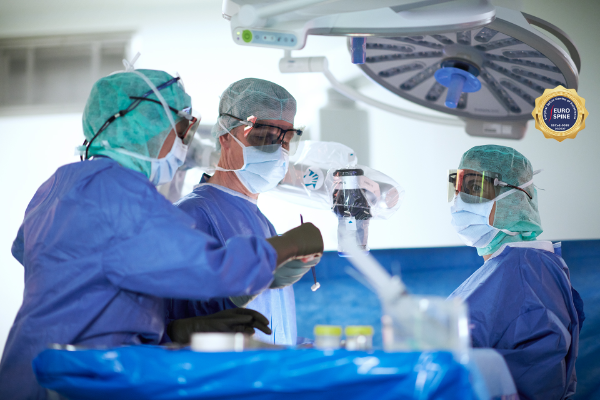
Spinal surgery comprises a team of spinal specialists from neurosurgery and orthopaedics. Their merger combines the knowledge and experience of both disciplines in a central specialist unit for patients with back problems. The interdisciplinary specialization within a large hospital allows us to precisely clarify even complex clinical pictures in order to treat their causes as gently as possible while preserving movement. As one of the largest spine departments in Switzerland, we are consulted around 13,000 times a year and carry out around 1,100 operations. We are active in clinical practice, research and teaching and hold the first structural professorship for spinal surgery in Switzerland.
Frequently asked questions about back pain
FAQ Back pain
What is spinal canal stenosis? - Prof. Stefan Schären
How does a herniated disc develop? - Dr. Morten-Goetz Wasner
How do osteoporotic vertebral body fractures occur? - Reinhold Full
What are the causes of spinal deformity/scoliosis? - PD Dr. Arne Mehrkens
What treatment options are there for spinal tumors? - PD Dr. Cordula Netzer
Radio reports
Consultation hours
Our consultation hours help our patients to gain a better understanding of spinal disorders and a treatment concept tailored to their needs. For this, we require a referral from a GP and, as a rule, prior imaging. During the consultation, we work with the patient to determine whether an operation is necessary and appropriate based on the available medical findings. Depending on the issue, other specialist disciplines such as rheumatology or pain therapy are included in the treatment chain.
Range of services
Wear and tear (degeneration), injuries, inflammation, tumors and malpositions can lead to changes in the spine that require surgical treatment. Problems with the spine can manifest themselves in pain, paralysis and/or other neurological disorders - such as impaired sensation, difficulty walking, bladder and bowel incontinence.
A prerequisite for the indication of surgery is a clear connection between your symptoms and the changes in the imaging. Not all changes that can be seen in X-ray, MRI or CT images should or must be treated surgically.
The field of spinal surgery covers the entire spectrum of spinal disorders.
Wear and degeneration
Many spinal disorders are caused by wear and tear. In addition to degenerative changes to the intervertebral discs (disc prolapse/disc hernia), these also include instability of the spine (spondylolisthesis = spondylolisthesis) and narrowing of the spinal canal (spinal canal stenosis).
Most of the changes mentioned above, which can be detected by X-ray or magnetic resonance imaging (MRI), are signs of normal age-related wear and tear. As long as there are no significant symptoms, no treatment is required. In the event of pain and discomfort, non-surgical therapies such as sports and exercise therapy, physiotherapy, medication and infiltrations (injections) can be used initially. In the case of complaints that do not improve spontaneously or through the above-mentioned therapies and if there are significant restrictions in everyday life, it can be examined whether there is a cause of pain that can be addressed by surgery.
Herniated disc
Intervertebral discs are like shock absorbers between the individual vertebrae and are therefore exposed to high mechanical stresses. There are various reasons why a so-called herniated disc, or disc prolapse, can occur. In this case, part of the disc bulges out or part of the soft core of the disc slips through the outer solid fibrous ring of the disc.
Not all people with a slipped disc have painful symptoms. However, if the part of the disc slips into an area where nerves run, these can come under pressure. In this case, pain, sensory disturbances, paralysis or even loss of bladder and rectal control can occur. Depending on the location of the herniated disc, different pain, discomfort or restrictions can result. The treatment options differ accordingly.
Lumbar spine
In the case of a herniated disc in the lumbar spine, the pain usually spreads down one leg, sometimes both legs, and part of the foot may also be affected.
A herniated disc is generally treated initially without surgery. However, surgical intervention may be necessary if symptoms persist and/or symptoms of paralysis occur. In this case, it is often sufficient to remove the part of the disc that is directly exerting pressure on the nerve. In very rare cases, it is necessary to remove the entire disc and replace it with an artificial prosthesis or to stiffen the entire segment at the same time.
The aim of such an operation is to prevent imminent damage to the nerve, to prevent the progression of nerve damage or even to improve disorders that have already occurred.
Operations on the intervertebral disc are usually minimally invasive and performed using a surgical microscope or endoscope.
Cervical spine
In the case of a herniated disc in the cervical spine, the pain typically spreads to the shoulder, arm or hand. Large prolapses can also lead to a narrowing of the entire spinal canal and pressure on the spinal cord, resulting in symptoms such as difficulty walking or impaired leg function.
In the case of minor complaints and restrictions, you can wait and see whether there is an improvement on its own. Physiotherapy, medication or injections can help to alleviate the symptoms. If the symptoms do not improve and there are restrictions in everyday life or even paralysis, an operation may be necessary.
In most cases, the entire intervertebral disc is removed from the cervical spine in order to relieve the nerve or spinal cord sufficiently. A placeholder is then inserted in place of the disc. There are two standard procedures:
- Stiffening of the segment to be treated
- Implantation of an intervertebral disc prosthesis
Which procedure is suitable in each individual case depends on various criteria and is decided on a case-by-case basis.
The aim of such an operation is to prevent imminent damage to the nerve, prevent the progression of nerve damage or even improve any disorders that have occurred.
Operations on the intervertebral disc are usually minimally invasive and performed using a surgical microscope.
Narrowing of the spinal canal
Spinal canal stenosis is a degenerative spinal disease with narrowing of the spinal canal due to wear and tear. Various structures are usually responsible for the narrowing, such as protrusions of the intervertebral discs, enlargement of the intervertebral joints and thickening of the ligaments. Narrowing of the spinal canal occurs most frequently in areas of the spine that are subject to heavy strain, i.e. in the middle to lower lumbar spine and sometimes also in the cervical spine.
In the affected segment, spinal canal stenosis sometimes leads to pain. The narrowing of the nerves often leads to an increase in pain when walking and standing, sometimes accompanied by a feeling of weakness in the legs and even paralysis. Typically, the walking distance becomes shorter and shorter and the symptoms can be quickly improved by sitting down or leaning the upper body forward.
If there is a narrowing of the spinal canal in the area of the cervical spine, this can also lead to weakness and numbness in the arms, radiating into the hands and fingers.
In mild cases of spinal canal stenosis, it can help to strengthen the back and abdominal muscles through consistent training. In the case of inflammatory changes, pain therapy with medication can also be used. Another option is the administration of infiltrations (injections) to individual nerves or into the spinal canal.
If the symptoms can no longer be controlled with these conservative measures, it should be checked whether there is a possibility of surgical treatment with the aim of relieving the nerves. This involves opening the canal surgically and removing constricting structures. In most cases, the operation is minimally invasive. Depending on the number of vertebral segments affected and the accompanying changes, additional stabilization or stiffening with a screw-rod implant may be advisable.
Instability of the spine
Spondylolisthesis is a condition in which a vertebra slips over an adjacent vertebra. This form of the disease can be congenital or acquired and usually affects the vertebrae of the lumbar spine. If the vertebra slips badly, nerves running between them can become constricted. This can lead to pain, numbness and weakness in one or both legs.
As long as the symptoms are tolerable and there is no nerve damage, the back and trunk muscles should be consistently trained to build up muscle strength. Medication can also be taken to control the pain. If the pain can no longer be controlled or symptoms of paralysis occur, it should be checked whether surgical stabilization of the spine is possible. In such an operation, the nerves are first relieved and then the unstable vertebrae are stabilized with a screw-rod system.
Poor posture and malposition
A distinction is made between poor posture and incorrect posture . Poor posture can be changed and improved through posture training and exercise. A malposition can no longer be improved by training alone due to a deformity or long-term incorrect posture with incorrect loads. Changes in the spinal column statics require treatment if they cause pain that permanently restricts and prevents everyday activities and if symptoms of paralysis develop or threaten to develop.
There are different causes for the development of a malalignment:
- idiopathic (without an identifiable cause)
- neuropathic (due to a nerve disease such as Parkinson's disease)
- degenerative (due to wear and tear)
- post-traumatic (after an injury)
- post-infectious (after an inflammation)
Depending on the type of curvature, a distinction is made between
- Scoliosis: Lateral S- or C-shaped curvature
- Kyphosis: hunchback, forward curvature of the spine
Depending on the cause, shape and extent of the change, we have various treatment options at our disposal:
- Regular independent training of the back and trunk muscles. Initially, this can also be done under physiotherapeutic guidance. In the medium and long term, training is then carried out independently and, above all, regularly.
- In the event of persistent restrictions and pain, the pain can be treated with medication depending on the origin of the pain. The possibility of additional treatment with injections (infiltrations) should also be examined.
In rare cases, it may also be necessary to perform surgery with the aim of correcting the incorrect posture. These are usually rather complex operations, for which the extent of the correction and the associated risks must be carefully discussed with the patient in advance.
Spinal neurosurgery
Spinal neurosurgery deals with the surgical treatment of rare spinal cord diseases and malformations of the spinal cord and its surrounding membranes, including
- Tumors of the spinal cord, nerves and spinal meninges (see spinal tumor surgery)
- Malformations of the spinal cord membranes and the spinal cord
- Cerebrospinal fluid loss syndromes
- Cavitation of the spinal cord
- Functional spinal neurosurgery
Malformation of the spinal cord membranes and the spinal cord
These congenital malformations are often summarized under the term spina bifida. However, other diseases such as idiopathic myelon herniation or tethered cord syndrome also fall under this malformation.
If surgery is necessary, it is performed microsurgically under neuropysiological monitoring (see above).
Cerebrospinal fluid loss syndromes
In rare cases, there may be a defect in the skin of the spinal cord. This can be congenital or caused by an accident, for example. Nerve fluid penetrates through this defect into the surrounding tissue and is absorbed there. The relative loss of volume of the cerebrospinal fluid results in the typical symptoms with position-dependent headaches (worse when standing/walking, better when lying down) and/or disturbance of the cranial nerves, often with muffled hearing or reverberation and occasionally double vision.
The aim is to close the defect after detecting it using special MRI examinations or myelography.
Initially, whenever possible, a minimally invasive attempt is made to close the leak by means of a blood patch (epidural injection of the patient's own blood). If this is repeatedly unsuccessful, the defect is closed in a microsurgical operation under neurophysiological monitoring (see above).
Cavitation of the spinal cord
A distinction is made between syringomyelia and hydromyelia. Syringomyelia is classically extrentric and chambered. Over time, there is an increase in size due to valve mechanisms. The pressure on the nerve tissue of the spinal cord increases, resulting in functional deficits in the sense of paraplegia.
Hydromyelia describes an enlargement of the central canal of the spinal cord and has no pathological significance.
Syringomyelia is caused by a disturbance of the cerebrospinal fluid circulation. The cause may be an adhesion of the subarchnoid space around the spinal cord or a tethered cord syndrome, often with a thickened and shortened filum terminale. A narrowing of the cerebrospinal fluid space, as in Arnold Chiari syndrome, can also result in the development of syringomyelia.
The adhesions in the subarachnoid space can occur after an accident, which is often initially accompanied by a spinal cord injury, an inflammation or without any recognizable cause. The formation of cavities can impair the function of the spinal cord and even lead to paraplegia. There is no conservative treatment option. The adhesions are surgically removed and the normal subarachnoid space is restored. By normalizing the flow of cerebrospinal fluid around the spinal cord, the syrinx often recedes. It is only rarely necessary to open the syrinx cavity, e.g. in the case of long-standing adhesions or adhesions that cannot be surgically removed. These procedures are also performed microsurgically under neurophysiological monitoring (see above).
A special variant of this disease that quickly becomes symptomatic is the subarachnoid web, which shows a typical image on MRI known as the scalpel sign.
Functional spinal neurosurgery
Functional spinal neurosurgery treats chronic pain syndromes and spastic movement disorders that cannot be adequately treated with medication or surgery. Our clinic has a well-established, close cooperation with other specialist departments and institutions:
- Pain therapy at the University Hospital Basel
- REHAB Basel Clinic for Neurorehabilitation and Paraplegiology
Functional spinal neurosurgery includes the following treatment options:
- Intrathecal drug application with implanted catheter/pump systems: Various drugs can be administered directly in the area around the spinal cord via an intrathecal catheter/pump system (including baclofen, opiates, clonidine). The aim is to achieve a significant dose reduction of the orally required medication and the associated side effects in patients with severe spasticity or severe therapy-resistant pain (e.g. tumor diseases). first, a catheter is inserted into the dural tube (= space filled with cerebrospinal fluid surrounding the spinal cord) via a lumbar puncture and connected to an external device. If the patient benefits from the intrathecal application of medication during the so-called test phase, the pump is implanted permanently in a second step.
- Therapeutic electrical stimulation of the spinal cord "spinal cord stimulators/ SCS (spinal cord stimulation)": Spinal cord stimulators are used to apply electricity directly to the spinal cord, which modulates pain processing and ultimately leads to a reduction in pain. The aim is to achieve an improvement in quality of life and a reduction in pain medication in patients with chronic pain and high levels of suffering who have been treated with medication and surgery. The clinical pictures include failed back surgery (chronic pain syndrome after back surgery(s) and CRPS (complex regional pain syndrome), and the first step is a detailed interdisciplinary evaluation by our colleagues in pain therapy and us. If there are suitable candidates, electrodes are then placed on the spinal cord on an outpatient basis under local anesthesia, led out of the skin via an extension and connected to a stimulator. This is followed by the so-called test phase (1-2 weeks). If the patient benefits from the stimulation during this time, the neurostimulator is implanted permanently.
- Diagnostic and therapeutic infiltrations (e.g. of nerve roots, facet joints, sacroiliac joint), radiofrequency ablation and thermocoagulation in cooperation with the in-house pain therapy clinic
Tumors and metastases
Primary tumor

Prof. Cordula Netzer, MBA
Leitende Ärztin Spinale Chirurgie
Leiterin Wirbelsäulenzentrum
Bone and soft tissue tumors
Spinal cord tumors

Dr. Morten Wasner
Stv. Chefarzt Spinale Chirurgie
Facharzt Neurochirurgie
Interdisziplinärer Schwerpunkt Wirbelsäulenchirurgie

Tumor consultation
We offer a separate consultation hour for our tumor patients. Due to the often great urgency, appointments can be made at short notice. If necessary, we will be happy to coordinate further clarifications and treatments with your family doctor or oncologist.
Accessibility
If you have urgent questions about spinal tumor surgery, please contact the duty physician for spinal surgery directly by phone or email:
Tel.
+41 61 265 78 30 (secretary's office)
+41 61 265 25 25 (Head Office)
Email:
spinalechirurgie@usb.ch
Tumor board
Tumors are always treated as a team. In order to ensure the best possible treatment, all patients with spinal tumors are discussed in the so-called "tumor board". This is an interdisciplinary panel of experts with specialists who have many years of experience in the treatment of tumor patients. More information
We will discuss the treatment recommendations and options in detail with those affected and respond to the patient's wishes and needs. If oncologists are already involved in the treatment, we will coordinate with them.
Spinal tumor surgery
Tumors of the spine
A distinction is made between tumors of ectodermal origin and tumors of mesodermal origin and metastases. Tumors of ectodermal origin are tumors of the spinal cord, nerves and spinal meninges. Tumors of mesodermal origin are primary tumors of the connective and supporting tissue of the spinal column.
Tumors of the spinal cord, nerves and spinal meninges
Very rarely, tumors can form in the area of the nerves, the spinal cord and the skin surrounding the spinal cord, which then affect the conductivity of the spinal cord and may even cause paralysis. Treatment is often surgical and highly standardized.
All patients are discussed on an interdisciplinary basis in a special tumor board consisting of specialists from radiology, oncology, neurology, neurosurgery, radiation oncology and neuropathology prior to surgery and the optimal treatment path is determined for the patient.
If an operation is necessary, it is carried out in accordance with the best possible safety standards. In addition to the microsurgical operating technique involving the surgical microscope, the functional monitoring of the spinal cord and nerves during the operation is also fully guaranteed under general anesthesia by means of neurophysiological monitoring using motor and somatosensory evoked potentials (MEP, SSEP) and electromyography (EMG). This offers the greatest possible success in preserving the function of the spinal cord in order to avoid paraplegia.
In Switzerland, the treatment of spinal cord tumors is classified as highly specialized medicine (HSM). It may only be carried out at clinics that can demonstrate a minimum annual number of cases and perform these procedures on a regular basis
Primary tumors of the spine
Primary tumors or first tumors have their origin in the spine itself and are extremely rare. In January 2018, Dr. Cordula Netzer published an article with the current state of knowledge according to international expert committees entitled "Treatment options for sarcomas of the spine". Treatment options for sarcomas of the spine
Metastases of the spine
Metastasis means tumor offshoot: the initial tumor is located in another organ or site and spreads from there to the spine. This can particularly be the case with prostate carcinoma, lung and breast carcinoma. Metastases can destroy parts of the spine and cause serious symptoms.
The diagnosis and treatment of tumors requires the cooperation of various specialists. If you are already being treated by a tumor specialist (oncologist), we will coordinate the further treatment concept with him or her or call in appropriate specialists for co-treatment and further treatment.
Surgical treatment of the spine is not necessary in every case, but may become necessary if:
- Severe pain leads to immobility.
- A vertebral fracture is imminent or has already occurred.
- Tumor-related constrictions of the spinal cord or individual nerves lead to paralysis or paraplegia.
If a vertebral body has to be removed due to a tumor or if it has been destroyed by a tumor, it may be necessary to replace this or several vertebral bodies with implants or the body's own bone chips. These are often major and complicated operations that require careful planning and follow-up treatment.
Therapy options
Depending on the type of tumor, the treatment of tumors can consist of three pillars:
- Chemotherapy and/or
- radiotherapy and/or
- surgery.
Further information: Oncology and radiation oncology
Surgery for tumors of the spine
At the University Hospital Basel, we treat our patients on the basis of the latest scientific findings and with state-of-the-art medical equipment. We therefore use all surgical techniques for tumors of the spine:
- Vertebroplasty/kyphoplasty
- decompression
- Stabilization: open or MIS (minimally invasive surgery)
- Enbloc resection/ reconstruction
- Radiofrequency ablation (thermal ablation) in collaboration with colleagues in interventional radiology
- Microembolization in collaboration with colleagues in interventional radioneurology
Furthermore, we use state-of-the-art implant materials such as carbon in the treatment of tumors, which can have advantages in the treatment of spinal tumors.
Spinal metastases
Referral information on spinal metastases
In order to optimize the treatment process, we have revised the referral modalities for you:
- If there is a suspicion of myelon compression or fracture due to a metastasis, clarification and, if necessary, treatment is required as quickly as possible.
- Once neurological deficits have occurred, they often increase within hours and the prognosis deteriorates rapidly.
- In your practice, it will generally not be possible to carry out the necessary clarifications promptly.
- Rapid referral of the patient can help to improve the treatment outcome.
Medical history
- Known carcinoma
- New onset or rapidly progressive pain in the area of the spine
- All types of pain are possible
- Mechanical pain (with osteolysis) - under stress
- Nociceptive pain (due to the tumor growth itself) - at night
- Neuropathic pain (due to compression of the nerves) - Radicular
Clinical examination findings
- New acute sensorimotor deficits/ radiculopathy
- Ataxia/ signs of myelopathy
- Conus-Cauda syndrome
Laboratory
- BB, inflammatory parameters (elevated in the context of tumor disease)
- DD Spondylodiscitis
Trauma
Fractures can occur in all areas of the spine. An appropriate treatment strategy must be selected depending on the cause of the fracture (accident, injury, osteoporosis, tumor) as well as the type and severity.
Not every vertebral fracture needs to be operated on. If there are options for healing and treatment without surgery, we will discuss this with you. In this case, it is important to have a short-term and often repeated check-up, usually with an X-ray, to ensure that the fracture is healing well. Immobilization with an adapted corset or neck collar is often essential during this time.
Surgical procedures to restore stability to the spine or to correct misalignments are carried out as an emergency or - if the situation allows - as planned operations. The surgical techniques available are as varied as the injuries themselves. Each fracture requires an individual decision regarding the best possible surgical technique.
Injuries can lead to narrowing of the spinal canal and/or instability of the spine. Depending on the injury pattern, the narrowing is repaired, the normal position of the spine is restored and sufficient stability is provided. Screw-rod implants are often used.
Under certain conditions, vertebral body fractures in osteoporotic bones can also be treated with a relatively minor operation. This involves filling cement into the vertebral body as part of a so-called vertebroplasty/kyphoplasty.
In any case, we will inform you in detail about surgical and non-surgical treatment options for fractures, as well as their advantages and disadvantages.
Infections
An infection caused by pathogens such as bacteria, viruses, fungi or parasites in the area of the spine, also known as spondylodiscitis or spondylitis, can be a life-threatening disease. It often manifests itself as sudden onset, very severe pain in individual areas of the spine. Typically, the pain is also present at rest and at night. These infections are often caused by the spread or distribution of pathogens in the body via the bloodstream and by the accumulation of pathogens in the area of individual vertebral bodies and/or intervertebral discs.
Here too, diagnosis is the first step before treatment. It is essential to identify the type of pathogen in order to be able to initiate appropriate treatment. If there are symptoms of loss or paralysis due to an abscess with pressure on the nerves, it may be necessary to perform emergency surgery.
The treatment concept, which often requires several months of antibiotic treatment, is developed in close cooperation with our infectious disease specialists.
Revision interventions
After spinal surgery, symptoms may occasionally recur. There are many different reasons for this, and further operations are sometimes necessary to correct them after extensive prior clarification. The reasons for so-called revision operations can be summarized in two categories:
- Complications immediately after an operation: secondary bleeding, infections, wound healing disorders.
- Late effects of a previous operation: For example, the so-called follow-up segment complaints after a fusion operation. In this case, symptoms recur after an initial operation. In this case, a very precise examination of the origin of the pain is required. Only when this has been confirmed can further options be discussed.
Methods
The following techniques and methods are used in spinal surgery.
- Invasive diagnostics (facet and root blocks, discography)
- Microsurgical/open decompression (cervical spine, lumbar spine)
- Transpedicular stabilization (open, percutaneous, dynamic)
- Interbody spondylodesis (ALIF, TLIF, XLIF)
- Vertebral body replacement for fractures/tumors (cervical spine, thoracic spine, lumbar spine)
- Intervertebral disc arthroplasty (cervical and lumbar)
- Vertebroplasty/kyphoplasty for osteoporosis fractures
Bewegungsempfehlungen
Egal ob Ihre Rückenschmerzen ohne Operation behandelt werden oder ob ein operativer Eingriff geplant ist – wir haben für beide Situationen hilfreiche Bewegungstipps für Sie zusammengestellt.
Bewegungsempfehlungen bei Rückenschmerzen
Bewegungsempfehlungen vor und während dem Spitalaufenthalt
Bewegungsempfehlungen bei Rückenschmerzen
Bewegungsempfehlungen vor und während dem Spitalaufenthalt