Offer
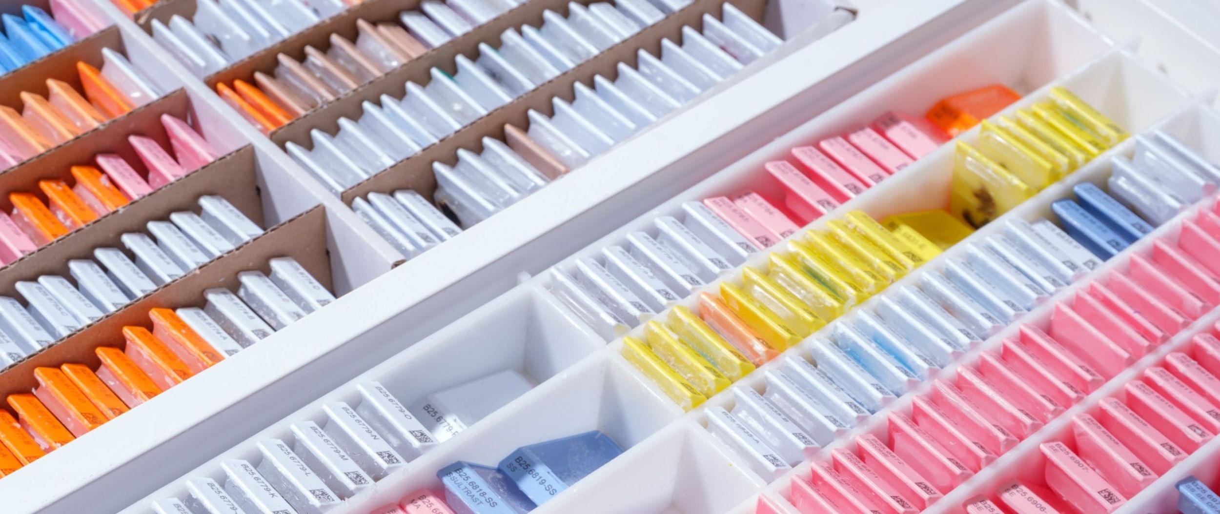
We examine patient samples using microscopy and molecular genetics to ensure that our diagnostics provide the basis for optimal treatment planning. Thanks to our close collaboration with the other clinics in the Department of Diagnostics and beyond, we are a central hub for highly specialized and academic medicine at the USB.
About us
As a university diagnostics service provider, we offer highly specialized diagnostics and genetic counselling for your patients throughout Switzerland. We - Laboratory Medicine, Pathology and Medical Genetics - work according to the motto "We work together for you" so that you can continue to concentrate fully on your patients. Personal support for digital order and result transmission tailored to your needs or comprehensive, coordinated sample logistics are just two of the many services you can expect from us.
Diagnostic procedures
The diagnostic spectrum of pathology includes the specialist areas of histopathology, cytopathology, molecular pathology and cytology, neuro- and ophthalmopathology, as well as post-mortem diagnostics (autopsy). The Institute is also home to the Bone Tumor Reference Center and the Reference Center for Malignant Lymphomas.
Continuous further training of all employees, the use of state-of-the-art technology and constant monitoring of work processes guarantee the highest quality in all areas. Pathology USB is accredited by the Swiss Accreditation Service SAS (SMTS 0037; ISO 15189:2012; SN EN ISO 15189:2013).
Histopathology
The histopathology department specializes in the examination of tissue samples from various organs. The aim is a high-quality macroscopic and microscopic assessment, which quickly leads to a correct diagnosis. The broad spectrum of techniques used includes special histochemical staining, enzyme and immunohistochemical examinations as well as special embedding and decalcification methods. Some tissue samples require a frozen section examination during an operation.
Biopsies are usually sent in standard fixative, 4% buffered formalin solution (can be obtained: pdf order form or phone +41 61 265 27 57), ideally in a ratio of at least 1:10 tissue sample to fixative solution.
For specific questions, it may be necessary to send non-fixed tissue.
For samples to be examined by electron microscopy, the standard fixative is 3% glutaraldehyde (can be obtained: pdf order form or phone +41 61 265 27 57).
Special shipping conditions apply for certain organ-specific examinations.
Frozen section examination
The frozen section examination is available for intraoperative diagnostics. Indications for a frozen section examination are questions about the dignity of lesions or completeness of the resection. It takes about 15 minutes (per sample, cumulative for several samples) from the arrival of the sample at the institute to the reporting of the findings.
The samples must be sent in their native state and dry (not in fixative or saline solutions). Findings are communicated by the pathologist to the sending clinic by telephone. It is therefore essential that a working telephone number at which the submitting person can be reached at the time of the frozen section examination is noted on the completed submission form.
The frozen section service is available throughout the Institute's opening hours (Mon-Fri 7.45-17.15). Pre-registration is required by calling +41 61 265 28 56.
For frozen section examinations that are required outside of these hours on weekday evenings, it is essential to make an appointment by telephone during the Institute's opening hours on +41 61 265 28 56.
The Institute provides telemedical support for the frozen section at the Hôpital du Jura in Delémont. Special IT requirements are necessary for this. This type of frozen section examination must be registered 24 hours in advance by e-mail(alexandar.tzankov@usb.ch or thomas.menter @usb.ch).
Dermatopathology
Indications for dermatohistological examination
- Unclear clinical pictures
- Chronic diseases that require prolonged therapy with many side effects
- Therapy and follow-up checks
- Medical-legal issues (expert opinions)
Clinical data
- Age and gender
- Medical history
- Previous topical or systemic therapies
- Clinical description (efflorescences and their distribution)
- Localization of the biopsy
- Sampling technique
- Previous biopsies
- Differential diagnosis
Biopsy sampling
- Unclear inflammatory skin diseases in various stages of development, polymorphic lesions or erythroderma: multiple biopsies.
- Suspected panniculitis, vasculitis or lymphoma: sufficient length (at least 2-3 cm) and depth of the specimen.
- Monomorphic lesions in disseminated distribution: one representative biopsy.
- Annular, centrifugally growing dermatitis, atrophies, ulcers and blistering skin diseases: Incisional biopsy at right angles to the marginal area with the center and periphery of the lesion including healthy skin.
- Leukocytoclastic vasculitis or bullous autoimmune dermatoses Direct immunofluorescence
Marking of biopsies
If an assessment of the resection margins is to be made for tumor excisions or post-resections, suture markings must be applied. Note the meaning of the suture markings on the submission form (e.g. suture at 12 o'clock, suture at the resection margin furthest from the tumor). A sketch is very helpful for more complex excisions
Prof. Katharina Glatz
Head of Pathology,
Member Tumor Center
Phone+41 61 328 68 94
kathrin.glatz@usb.ch
Bone marrow
Bone marrow biopsies
In order to enable a standardized diagnosis of bone marrow biopsies, we ask the referring clinical colleagues to provide the following information:
- Clinical (suspected) diagnosis,
- Reason for taking the biopsy (e.g. lymphoma staging, clarification of leukocytosis, pancytopenia, etc.),
- Current blood count with differential blood count,
- Information about:
- known (hematologic) neoplasms, including chemo/radio/immunotherapies performed,
- other systemic diseases (e.g. granulomatous processes, autoimmune diseases, storage diseases, etc.),
- current and/or previous, in particular myelotoxic/myelosuppressive therapies (e.g. azathioprine, arsenic, etc.), exposure to hematopoietic growth factors (erythropoietin, G-CSF, interferon, etc.),
- previous autologous/syngeneic or allogeneic stem cell transplants or organ transplants,
- acute/chronic viral diseases (CMV, EBV, HBV, HCV, HIV, Parvo-B19, etc.) or any other infections,
- Presence of organomegaly,
- other relevant findings/information (e.g. results of aspiration cytology, flow/FACS analyses, cytogenetics, etc.),
- if possible, information on vitamin B12, folic acid and iron status.
The bone marrow biopsies must be sent in 4% buffered formalin solution (can be obtained: order form or phone +41 61 265 27 57).
Please use the submission form for bone marrow biopsies.
Contact for medical questions
Prof. Stefan Dirnhofer
Deputy Head of Pathology
Member Tumor Center
Phone+41 61 328 67 89
stefan.dirnhofer@usb.ch
Prof. Alexandar Tzankov
Head of the Department of Histopathology and Autopsy
Member Tumor Center
Phone+41 61 328 68 80
alexandar.tzankov@usb.ch
Paidopathology and placenta diagnostics
Solid tumors in childhood differ morphologically and molecularly as well as therapeutically and prognostically from those in adulthood. Depending on the entity, they therefore require special processing with various stains and methods available at our institute, including molecular pathological examinations such as fluorescence in situ hybridization (FISH) and mutation analyses with specialized next generation sequencing (NGS) panels.
This results in the following recommendations for our senders: To enable these examinations, tumor biopsies and tumor resectates should be sent in unfixed, fresh, native, on water ice by courier after prior notification by telephone. In close collaboration with the University Children's Hospital Basel (UKBB), we ensure the participation of patients in studies of the Swiss Pediatric Oncology Group (SPOG) and international study consortia by sending sample material to the appropriate reference centers. Furthermore, a routine reference assessment is carried out by the Children's Tumor Registry in Bonn (Prof. Christian Vokuhl) in consultation with the UKBB Oncology Department.
The histopathological assessment of placenta and abortion curettage can make an important contribution to understanding fetal and maternal pathologies and also to clarifying the cause of death in the event of intrauterine fetal death. Information on the week of pregnancy, findings of the child and the mother as well as any abnormalities during pregnancy and the birth process are helpful for an adequate assessment. The tissue can normally be sent in formalin-fixed, but if the fetus is to be assessed as well as the placenta, it should be sent in unfixed as soon as possible after birth.
Contact us
PD Dr. Thomas Menter
Head Physician Pathology
Phone+41 61 328 68 97
thomas.menter@usb.ch
Motility disorders, Hirschsprung's disease
The diagnosis of aganglionosis in Hirschsprung's disease can be made reliably on the basis of frozen sections using enzyme histochemical imaging of the increased acetylcholinesterase activity of the parasympathetic nerve fibers of the muscularis mucosae and the lamina propria of the rectum as well as the lack of evidence of ganglia.
Hypoganglionosis is typically found in the proximal segment preceding the aganglionic segment, the extent of which should be recognized in order to perform the resection in a functionally intact section of bowel. The enzyme histochemical reaction for lactate dehydrogenase (which can also be carried out intraoperatively on the frozen section) is able to differentiate this.
Close cooperation with the Intest Team of the University Children's Hospital Basel (UKBB), which deals with intestinal motility disorders in children and adolescents, enables interdisciplinary care of patients.
Instructions for submitting tissue for examination for Hirschsprung's disease: Hirschsprung's disease material submission
Instructions for the submission of sectional and kerosene material for consultation pathology assessment: Consultation pathology assessment
Contact for medical questions

Prof. Alexandar Tzankov
Leitender Arzt und Fachbereichsleiter Histopathologie und Autopsie
Pathologie
Mitglied Tumorzentrum
Tel. +41 61 328 68 80

Laboratory: Tel. +41 61 265 28 56
Fax +41 61 265 31 94
Cytopathology
The specialist area of cytopathology examines changes in body cells that are obtained from body surfaces, fluids and organs, processed in the laboratory, assessed under the microscope and, if necessary, examined using additional methods. Diseases and changes in practically all organs can thus be diagnosed easily and reliably.
Gynecological screening cytology is used for the early detection of precursor lesions of cervical cancer. The liquid-based method guarantees optimum specimen quality and, if required, a delay-free HPV examination. We use the currently valid algorithms of the Swiss Society of Gynecology and Obstetrics for control recommendations in the event of abnormal findings.
In extragenital cytology, the focus is on the diagnosis of tumors with examination of prognostic and predictive markers. This is done using the latest techniques. Detailed information can be found under Molecular Pathology and Cytology.
Special methods are used for specific issues:
- Immunocytochemistry: diagnosis and classification of malignant tumors as well as investigation of prognostic and predictive markers
- Immunofluorescence: pathogen diagnostics in bronchoalveolar lavages
- Fluorescence in-situ hybridization (FISH): improved tumour diagnostics and predictive marker analyses
- Laser microdissection: enrichment of tumor cells for PCR-based DNA sequencing
-
Certain tests require special shipping conditions:
Gynecological examination
1. gynecological smear test
Liquid-based gynecological smears (ThinPrep® method):
- With the ThinPrep® method, lubricant gel can lead to clumping of the cells with limited assessability of the preparations. However, if necessary, a small amount of a water-soluble carbomer-free lubricant can be used (e.g. Gyn-Lys or K-Y lubricant medical Steril).
- Rinse the collection brush and/or spatula well in the PreservCyt® solution and rotate along the wall or bottom of the container to detach the cells well.
- Then dispose of the brush or spatula (do not leave in the solution).
- Send the container.
Conventional (classic) gynecological smears:
- Use slides with a matt end piece and label with the patient's surname, first name and date of birth in pencil.
- Spread the cell material quickly (within 2-3 seconds), thinly and evenly in one movement by rolling the brush or swabbing the spatula (no cotton swabs).
- and fix immediately: spray with fixation spray until the preparation is completely covered by a liquid film. Allow smears to dry for 10-20 minutes before shipping.
- Dispatch in shipping tubes.
2. HPV typing
Typical indications:
- Unclear cytologic findings (ASC-US, ASC-H and AGC) in patients ≥ 30 years.
- Mild dysplasia (Bethesda low grade SIL) in patients ≥ 30 years of age
- Primary HPV screening
Material collection for HPV examination:
- Liquid cytology: HPV determination on the remaining residual fluid ("reflex HPV testing"). Residual material from liquid cytology is available for 4 weeks for subsequent HPV testing.
- Conventional smears: Test kit (Digene Specimen Collection Kit, QIAGEN) with collection instrument (provided by us). Material collection on the occasion of a control examination.
3. endometrial aspirates
Material collection:
- Aspiration of the uterine cavity with a balloon pipette
- Collection of the aspirate in AMIMED® liquid medium
Fine needle puncture
Important notes
Have all utensils ready before the fine needle aspiration (FNP).
Check the function of the fixation spray before the puncture. Only use slides that can be labeled (surname, first name and date of birth in pencil. No felt-tip pens, no labels). Label before smearing the cell material.
If lymphoma is suspected (lymph node FNP), prepare additional punctate for FACS analysis in hematology.
Fixation
- Fix smears immediately after puncture (within seconds!)
- Spray fixation: spray at a distance of approx. 10-20 cm until the specimen is completely covered by a liquid film. Allow smears to dry for 10-20 minutes before shipping.
or - Wet fixation using Delaunay solution (can be ordered from the cytopathology laboratory on +41 61 556 53 66).
Shipping
- Spray-fixed: Shipping tubes
- Moist-fixed (Delaunay solution): Shipping container with screw cap
- Rinse FNP syringe with 0.9% NaCl or cell medium after smearing and send with rinsing liquid
- If lymphoma is suspected, send separate punctate directly to the hematology laboratory
Technique of fine needle aspiration (FNP)
Series of online videos with detailed discussion of the FNP technique:
Bronchoalveolar lavage (BAL)
Indications
- Cell differentiation with determination of the CD4/CD8 ratio ("immune lavage") for suspected inflammatory or interstitial lung disease
- Determination of lymphocyte subpopulations (CD4, CD8, CD3 and CD19) using FACS
- Clarification of infection ("infection lavage")
- Pathogen detection using special tests (immunofluorescence, special stains): Pneumocystis jirovecii (PCP), CMV, RSV, Legionella pneumophila, mycobacteria, fungi
Registration
- If urgent diagnosis is required on the same day: please register BAL with the laboratory (tel. +41 61 556 53 66) at the latest immediately before the start of the procedure and state the problem (cell differentiation/search for infection/malignancy).
- Urgent clarification of infection by means of special tests: Results on the same day, provided BAL arrives at the cytopathology laboratory by 2 p.m. at the latest.
Preservation
- No preservatives. Transport on ice water (water/ice in equal parts). Do not place the container in pure ice.
Shipping
- In graduated plastic container with direct transport within the hospital (STA until 5 p.m., thereafter in consultation with cytopathology laboratory): For postal or rail transportation (express or courier service) in a watertight sealable plastic container.
Endobronchial examination
Sputum
Indications
- If a tumor is suspected, collection of fasting sputum on three consecutive days (sputum I-III).
Procedure
- Brush teeth before sputum collection.
- Denture wearers: remove dentures, rinse mouth well.
- Inhale deeply and cough up deep-seated mucus.
- If there is no sputum, inhale with 3% saline solution and tap on the back over the diseased lung, causing viscous bronchial secretions to vibrate and triggering a cough.
- Spit the coughed up mucus directly into a container intended for sputum or into a sputum cup.
Preservation
- For transportation times of less than 1 day: no addition.
Shipping
- Send each sputum sample immediately and separately. Do not collect sputum.
- Send in tightly sealable plastic containers with a wide opening.
Outpourings
The following applies to all serous fluids (pleural and pericardial effusion, ascites, aspirate from Douglas's space, peritoneal lavage):
- No addition of preservatives. Sterile serous fluids are good
- good cell media. Cells can be stored at room temperature for approx. 24 hours, at 4 °C in the refrigerator over the weekend.
- Send the entire volume of fluid (at least 1 liter for large volumes, preferably all of it)
Urinary tract cytology
Spontaneous urine
Procedure
- First empty the bladder on the morning of the examination
- Then drink several glasses of liquid
- Exercise before the 2nd bladder emptying (gymnastics, climbing stairs, walking)
- Empty the bladder completely into the container provided
- Women: Dab labia with a wet cloth before micturition (avoid genital squamous epithelia in the urine)
Preservation
- Immediately add 50% alcohol in a 1:1 ratio to the urine sample (to preserve and suppress bacterial growth).
Dispatch
- Send the sample immediately so that it can be processed on the same or next day if possible.
Catheter urine
Like spontaneous urine, immediately mix with 50 % alcohol in a 1:1 ratio.
Important: Note on the submission form whether it is spontaneous or catheter urine.
Urinary bladder irrigation fluid
Preservation
- Immediately add 50 % alcohol in a 1:1 ratio (as for urine).
UroVysion™ FISH
Examination of urothelia for chromosomal aberrations. We offer you this examination for further clarification depending on the cytological findings. It is performed on the cytology specimen that has already been prepared and does not require the separate submission of additional material.
Most important indications:
- Unclear cytological atypia (e.g. upper urinary tract, after BCG or as part of follow-up care)
- Discrepancy between cytology and cystoscopy
Cerebrospinal fluid
- If cerebrospinal fluid is to be examined exclusively for tumor cells, please send it directly to the cytopathology laboratory in the pathology department.
- In all other cases, send cerebrospinal fluid to the Laboratory Medicine Department of the University Hospital Basel, where cell count, protein, glucose etc. are determined. From there, cytocentrifuge preparations are forwarded to the cytopathology laboratory for cell differentiation.
- Cerebrospinal fluid is a nutrient-poor fluid. The cells therefore die quickly. In addition, cell adhesion to the walls of the sending vessel leads to cell loss. This results in
- CSF must be processed within 2 hours and sent to the laboratory immediately.
Preservation
- Only native CSF without added preservatives
Shipping
- Do not send too small a quantity (≥ 2 ml if possible)
- Send in a plastic tube (cell adhesion lower than in glass tubes)
Immunocytochemistry
A wide range of antibodies is available for immunocytochemical testing of neoplastic and non-neoplastic diseases (for tumor typing, prognostic and predictive marker tests and pathogen diagnostics). Immunocytochemistry is performed directly on the diagnostic cytological preparations or on cell blocks.
Molecular pathology and cytology
Molecular pathology and cytology includes molecular pathological and molecular genetic examination methods. These include in-situ methods (e.g. fluorescence in-situ hybridization (FISH), immunohistochemistry and immunocytochemistry), PCR-based methods (DNA, RNA) and next generation sequencing (NGS). These methods support the treating physicians in choosing the right therapy and provide important information on prognosis and prediction (e.g. response to medication), particularly in the case of tumor diseases.
The most modern examination methods are available to you for the various diagnostic, prognostic and predictive questions.
Fluorescence in situ hybridization (FISH)
This is a method that can be used for both unfixed and fixed material. For this purpose, section/block preparations or cytological preparations (also already stained) can be used. The principle is based on the hybridization of gene probes to the gene sections of the test material to be examined. These gene probes are coupled with fluorescent dyes that can be visualized under a special fluorescence microscope.
Our range of analyses includes a number of different probes, which are used in particular for the diagnosis of lung carcinomas, sarcomas and lymphomas. Numerical genetic aberrations (amplifications and deletions) as well as translocations can also be detected.
This method is also suitable for clarifying unclear changes in cytology (e.g. Urovision® for changes in the urinary tract.
Please use the following forms for submission:
Next Generation Sequencing (NGS)
Next Generation Sequencing (NGS) makes it possible to obtain an overview of numerous clinically relevant gene alterations (e.g. point mutations, deletions, insertions and copy number changes) in tumor tissue with a single analysis. The method makes it possible to sequence many different DNA sequences simultaneously and thus obtain information on numerous therapeutically relevant genes at once.
Commercially available kits from Thermo Fisher and ArcherDx as well as panels developed in-house are used for this purpose.
DNA-based analyses are carried out with the following panels, among others:
- Oncomine™ Precision Assay GX (primarily tumors of the lung, colon, breast, GIST), 46 genes with 14 copy number variations
- CE-IVD Oncomine™Solid Panel (mainly tumors of the lung and colon; 22 genes)
- Oncomine™ Comprehensive Assay v3 (extended gene spectrum for all tumors, 135 genes, 47 copy number variations)
- Oncomine™ Comprehensive Assay Plus (including TMB) (extended gene spectrum for all tumors, including tumor mutation burden, 498 genes with 333 copy number variations)
- Oncomine™ Childhood Panel (primarily childhood tumors including DICER1 and MYOD1), 136 genes with 28 copy number variations
- HRR-BRCAness Prostate Panel (mainly tumors of the ovary, prostate, breast and pancreas) with BRCA1/2, RAD51B, ATM, CDK12 and other genes, 55 genes
- Lymphoma panel (frequent mutations in a total of 68 lymphoma genes)
- Melanoma panel (frequent mutations of melanoma) with BRAF, NRAS, KIT, TERT, NF1, GNAQ, GNA11, BAP1 and other genes, 33 genes
In addition, we also offer RNA-based analyses that can be used to detect gene fusions of e.g. ALK, ROS1, RET, NTRK1/2/3 and FGFR2/3. These include:
- Archer™ FusionPlex™ (fusions of 137 genes)
- Oncomine™ Comprehensive Assay v3 RNA (fusions of 51 genes)
- Oncomine™ Precision Assay GX (fusions of 18 genes)
- Oncomine™ Focus Assay RNA (fusions of 23 genes)
- Oncomine™ Childhood RNA Panel (fusions of 91 genes)
We also offer the determination of tumor mutation burden (TMB). This procedure uses NGS to determine the number of mutations per 1 million base pairs. Determining the TMB can help to identify patients who are more likely to benefit from immunotherapy.
In principle, NGS methods can be used for both histological and cytological specimens. Using laser capture microdissection, we can increase the tumor cell content so that evaluable results can be obtained even with few tumor cells or a low tumor cell content.
A detailed list of analyzable genes and exons as well as further information on NGS can be found here:
Liquid biopsy
We offer the examination of cell-free DNA (cfDNA) in peripheral blood and cerebrospinal fluid under the name liquid biopsy. This fragmented DNA, which originates from numerous tissues of the body, also contains so-called circulating tumor DNA (ctDNA) in tumor patients. Appropriately sensitive methods can be used to search for therapy-relevant mutations, in particular those responsible for drug resistance. Another common application is the quantification of ctDNA based on already known tumor-specific mutations that were previously determined in conventional biopsies. In follow-up measurements, an increase in ctDNA in the peripheral blood usually indicates tumor progression.
The analysis is usually performed with an NGS method using various panels such as Oncomine™ Lung cfTNA, Oncomine™ Colon cfDNA Assay, Oncomine™ Breast cfDNA Assay or Oncomine™ PanCancer cfTNA Assay. These panels can be performed generally for various malignancies, or specifically for malignancies of the lung, colon and breast.
As ctDNA must be protected from dilution by leukocyte DNA, which is released by cell decay during storage of blood samples, prepared vacutainers with preservative (e.g. Streck tubes) must be used. A blood sample or cerebrospinal fluid sample can be stored and sent in these containers for approx. 4 days at room temperature. 20 ml of blood is required for one examination, if possible 2 ml of cerebrospinal fluid should be sent in. Appropriate tubes can be ordered from us on request (Tel: +41 61 265 27 57).
Central to the liquid biopsy are the clinical question and the most detailed information possible on the confirmed or suspected tumor in order to obtain the maximum amount of clinically usable information from the sample. This information should be provided on the submission form.
The corresponding submission form and a detailed list of analyzable genes and exons can be found here:
DNA methylation
We offer array-based genome-wide DNA methylation analyses for tumor precision diagnostics (1,2). These analyses are available for brain and soft tissue tumors, peripheral nerve sheath tumors, and in particular for CUP syndrome (cancer of unknown primary) (1,3). For CUP diagnostics, the data sets are matched against the DNA methylation profiles of more than 18,000 tumors using the in-house developed EpiDiP system (4), which in most cases allows assignment to a primary or tissue of origin.
In addition totumor entity-specific DNA methylation profilesmicroarray analysis provides a wealth of other important information:
- Copy number profiles, including LOH 1p/19q in gliomas, MDM2 gene amplification in adipose tissue tumors
- Prognostic information, e.g. CDKN2a/b deletions in IDH-mutated gliomas, molecular meningioma grading (5), MLH1 promoter methylation
- Predictive markers, e.g. MGMT promoter methylation, EGFR and HER2 gene amplifications
Prerequisite for the analysis is 500 ng DNA (preferably from native tissue, but FFPE is also possible).
We also offer a molecular rapid test based on nanopore sequencing (6,7), which allows reliable methylome classification in just a few hours using the NanoDx (7,8) and NanoDiP (9) algorithms. This analysis is reserved exclusively for native specimens and specimens preserved using SurePath® or ThinPrep® (including malignant effusions). The preservation of native tissue samples in SurePath® or ThinPrep® allows external submissions at room temperature. Following the rapid test, the findings are verified using a high-resolution microarray (takes approx. 2 weeks). Please use the following forms for submissions:
References
1.Capper D, Jones DTW, Sill M, Hovestadt V, Schrimpf D, Sturm D, et al. DNA methylation-based classification of central nervous system tumors. Nature. 2018 Mar 14;555(7697):469-74.
2.Hench J, Bihl M, Bratic Hench I, Hoffmann P, Tolnay M, Bösch Al Jadooa N, et al. Satisfying your neuro-oncologist: a fast approach to routine molecular glioma diagnostics. Neuro-Oncol. 2018 Nov 12;20(12):1682-
3.Koelsche C, Schrimpf D, Stichel D, Sill M, Sahm F, Reuss DE, et al. Sarcoma classification by DNA methylation profiling. Nat Commun. 2021 Dec;12(1):498.
4.Hench J, Frank S. EpiDiP Server [Internet]. 2020. available from: http://www.epidip.org
5.Maas SLN, Stichel D, Hielscher T, Sievers P, Berghoff AS, Schrimpf D, et al. Integrated Molecular-Morphologic Meningioma Classification: A Multicenter Retrospective Analysis, Retrospectively and Prospectively Validated. J Clin Oncol. 2021 Dec 1;39(34):3839-52.
6.Euskirchen P, Bielle F, Labreche K, Kloosterman WP, Rosenberg S, Daniau M, et al. Same-day genomic and epigenomic diagnosis of brain tumors using real-time nanopore sequencing. Acta Neuropathol (Berl). 2017;134(5):691-703.
7.Kuschel LP, Hench J, Frank S, Hench IB, Girard E, Blanluet M, et al. Robust methylation-based classification of brain tumors using nanopore sequencing [Internet]. Oncology; 2021 Mar [cited 2021 Apr 1]. Available from: http://medrxiv.org/lookup/doi/10.1101/2021.03.06.21252627
8.Djirackor L, Halldorsson S, Niehusmann P, Leske H, Capper D, Kuschel LP, et al. Intraoperative DNA methylation classification of brain tumors impacts neurosurgical strategy. Neuro-Oncol Adv. 2021 Oct 10;vdab149.
9.Hench J, Hultschig C. NanoDiP - Nanopore Digital Pathology [Internet]. NanoDiP - Nanopore Digital Pathology. 2021 [cited 2021 Nov 26]. Available from: https://github.com/neuropathbasel/nanodip
contact
Prof. Stephan Frank
Head of the Department of Neuro- and Ophthalmopathology, Member of the Tumor Center
Phone+41 61 328 63 90
stephan.frank@usb.ch
Dr. Jürgen Hench
Senior Physician Neuro- and Molecular Pathology, Member Tumor Center
41 61 328 68 91
juergen.hench@usb.ch
Other methods
Sanger sequencing
Sanger sequencing after PCR amplification of DNA is used to search for mutations within defined genome sections. The Sanger reaction products are analyzed by capillary electrophoresis, an improvement on the polyacrylamide gel electrophoresis initially used by Sanger. At the time, this method was instrumental in enabling and shaping the sequencing of entire genomes before it was largely replaced by parallel sequencing (also known as next generation sequencing, NGS). Nevertheless, the Sanger technique is still the reference standard for gene sequencing today and is used to validate new technologies. We still use Sanger sequencing primarily as a cost-effective method for determining mutations in individual gene regions.
Fragment length analysis using capillary electrophoresis
Like Sanger sequencing, this method is based on the capillary electrophoretic separation of specifically generated PCR products from sample DNA and their length determination. Depending on the PCR approach, the technique can be used for clonality analysis in lymphoproliferative diseases or to determine microsatellite instability in carcinomas.
Droplet digital PCR
In droplet digital PCR (ddPCR), the DNA sample is divided into manydroplets (up to 20,000 per reaction) in a water-oil emulsion. Subsequently, a mutation-specific PCR is performed in each of these individual droplets using fluorescence-coupled TaqMan probes. All droplets are then analyzed for the presence of a mutation, with those in which mutation-specific amplification has occurred fluorescing. The analysis is carried out using a modified fluorescence flow cytometer. As a rule, two dyes are used to distinguish the mutant allele from the wild-type allele. The ratio of the two color signals corresponds to the allele frequency of the mutation (number of mutated alleles per total number of alleles in the tumor tissue). In contrast to the Sanger method, ddPCR is a very robust method for searching for single-gene mutations, as the allele frequency is always determined, which makes it easier to interpret the findings. Due to the quantitative results, ddPCR can also be used to determine gene amplifications, deletions and DNA methylation.
Human papillomavirus (HPV) genotyping
Genotyping is necessary to determine whether an HPV infection exists and whether this is associated with an increased risk of progression or carcinoma development. A quantitative PCR method is used for this purpose, which not only differentiates between high risk and low risk HPV genotypes, but in the vast majority of cases also determines the exact HPV types involved (even in the case of multiple infections). The method can be used for all cytological procedures, but also for formalin-fixed and paraffin-embedded and native tissue. This analysis is carried out in collaboration with the Clinical Virology Department of Laboratory Medicine at the University Hospital Basel.
The corresponding submission form can be found here:
Contact for medical questions

Prof. Lukas Bubendorf
Leitender Arzt und Fachbereichsleiter Zytopathologie
Pathologie
Tel. +41 61 328 78 51

Prof. Matthias Matter
Leitender Arzt und Fachbereichsleiter Molekularpathologie
Pathologie
Tel. +41 61 328 64 71
Information on molecular tests

Neuro- and ophthalmopathology
The Department of Neuropathology deals with the diagnosis of diseases of the central and peripheral nervous system, including diseases of the skeletal muscles (myopathology). Our focus is on the molecular analysis of a broad spectrum of tumor diseases, including brain tumors, using DNA methylation profiling; we also use the latest nanopore sequencing methods and various artificial intelligence algorithms. Another focus is on the diagnosis of nerve and muscle diseases. In addition, we carry out examinations on the brains of deceased patients, with a particular focus on the diagnosis of neurodegenerative diseases and fetal brain malformations (collaboration with Medical Genetics, USB).
The Department of Ophthalmopathology focuses on the histological examination of surgically removed eye tissue (eyelids, conjunctiva, cornea, orbit, eyes). This service is provided for the USB Eye Clinic, for external ophthalmology departments at acute hospitals and eye clinics as well as for independent ophthalmologists. In addition to specific histotechnical procedures, molecular pathological examination methods are increasingly being used.
Clinical information for muscle and nerve biopsies
Muscle biopsy
In order to ensure professional histological processing of the native sample, advance notification is required either by e-mail or by telephone +41 61 328 78 74. Whenever possible, this pre-registration should be made at least 24 hours before the biopsy is taken.
Send muscle tissue fragment (size approx. 1x1x1 cm) native (unfixed, without medium) in a closed container (e.g. thermos, polystyrene box) on ice by cab, courier, etc. Caution: Tissue not in direct contact with ice. Do not use dry ice or frozen ice packs!
If cab or courier transport is not possible, please send biopsy specimen by express mail (same-day delivery, schedule biopsy accordingly) to:
Pathology
University Hospital Basel
Schönbeinstrasse 40
4031 Basel
Within the University Hospital Basel: pneumatic tube 2856, please call 87874 when sending.
In general
- Please always indicate where the muscle was taken.
- Please enclose a medical report with the biopsy specimen, stating clinical symptoms and questions in as much detail as possible. Name the medical contact person.
- The biopsied muscle should be affected by the symptoms of the disease described, but if possible it should not be clearly paretic/plegic or remodeled muscle.
- EMG needles cause considerable artifacts. Therefore, do not take muscle biopsies from areas into which EMG needles have been inserted.
Clinical information form for muscle/nerve biopsies
Nerve biopsy
Pre-registration of the nerve biopsy either by e-mail or by telephone +41 61 328 78 74. Whenever possible, pre-registration should be made at least 24 hours before the biopsy is taken.
Nerve fragment at least 2 cm long, ideally 4 cm long (in children not less than 1 cm if possible).
Within the University Hospital Basel: Send in native condition by pneumatic tube 2856. If sending, please call 87874.
Other senders: either fixed in 4% buffered formalin (regular mail) or cooled native by courier (see muscle biopsy).
Clinical information form for muscle/nerve biopsies
Registration/administration/order: Tel. +41 61 328 78 74
Contact us
Prof. Stephan Frank
Head of the Department of Neuro- and Ophthalmopathology, Member of the Tumor Center
Phone+41 61 328 63 90
stephan.frank@usb.ch
Dr. Jürgen Hench
Senior Physician Neuro- and Molecular Pathology, Member Tumor Center Tel.+41 61 328 68 91
juergen.hench@usb.ch
Contact us

Prof. Stephan Frank
Leitender Arzt und Fachbereichsleiter Neuro- und Ophthalmopathologie
Pathologie
Klinische Professur für Neuro- und Muskelpathologie
Tel. +41 61 328 63 90
Prof. Peter Meyer
Leitender Arzt
Augenklinik
Facharzt Ophthalmologie und Ophthalmochirurgie
Post-mortem diagnostics
The main task of post-mortem diagnostics (autopsy) is to determine the cause of death and pre-existing illnesses of a deceased patient by means of an internal medical post-mortem examination with specimen and - if necessary - organ autopsy. Pathological-anatomical (clinical) autopsies are carried out on people who have died of natural causes (otherwise forensic autopsies), provided they or their next of kin have given their consent.
The autopsy also serves
- quality assurance and quality control in clinical medicine,
- teaching (training of medical students, assistant doctors, specialist pathologists and specialists from other disciplines),
- answering questions from relatives, such as information on familial risk factors for cancer or dementia,
- research,
- clarifying insurance issues, for example in the case of occupational illnesses.
Contact us

Prof. Alexandar Tzankov
Leitender Arzt und Fachbereichsleiter Histopathologie und Autopsie
Pathologie
Mitglied Tumorzentrum
Tel. +41 61 328 68 80

Reference centers
The expertise for mostly rare diseases is concentrated in medical reference centers. In pathology, reference centers often take on the role of a second opinion, possibly also for inclusion in clinical studies, and have the relevant expertise and a specific technical infrastructure for additional examinations (e.g. gene sequencing). The reference pathological assessment of tissue samples from rare diseases enables a rapid, precise and reliable diagnosis and prognosis assessment and is the prerequisite for the initiation of optimal and "targeted" therapy.
Switzerland's only reference centers for bone tumors and lymphomas, whose positive reputation extends far beyond the country's borders and represents a beacon function for the USB, are located in the pathology department of the USB.
Bone tumor reference center
Bone Tumor Reference Center (KTRZ) and DÖSAK Reference Register
Bone and jaw tumors are rare overall, with malignant forms accounting for only approx. 0.5% of all malignant tumors (approx. 60 cases/year in Switzerland). Whereas in the past most patients died from these diseases, today 5-year survival rates of up to 70% are possible, especially for the highly aggressive forms that occur mainly in children and adolescents (e.g. osteosarcoma, Ewing's sarcoma). The prerequisite for this is a precise and reliable diagnosis, which requires a concentration of expertise in view of the numerous different subtypes with varying prognoses.
The Bone Tumor Reference Center and the reference register of the DÖSAK (German-Austrian-Swiss Working Group for Jaw and Facial Tumors) at the USB in Basel examines almost 1000 cases from Switzerland and abroad every year and enjoys an excellent reputation. The Bone Tumor Reference Center and DÖSAK Reference Registry was founded in 1972 on the initiative of Prof. Dr. H.U. Zollinger, former head of the Institute of Pathology, and was headed by Prof. Dr. W. Remagen until 1993 and by Prof. Dr. G. Jundt until June 2014. Prof. Dr. Daniel Baumhoer has headed the reference center since July 2014. The good cooperation with the clinics and tumor centers at the USB and UKBB has promoted and supported the clinical focus in Basel and attracts patients from all over Switzerland and neighboring countries.
The tasks of the reference center, which is supported by a foundation, consist of primary diagnostics and consultative second opinions. Since its establishment, more than 20,000 bone tumors have been examined and documented at the reference center, with particular attention paid to the inclusion of corresponding imaging and the clinical course. In around 40% of cases, the diagnoses of external submissions are changed and/or clarified; in 5% of cases, reclassifications from benign to malignant or from malignant to benign have to be made.
The documentation of the bone tumors examined at the reference center is the basis for precise diagnostics, advanced training events for pathologists, radiologists, orthopaedists and oral surgeons, as well as the basis for numerous publications and books. Particularly noteworthy is the co-design of the current WHO classifications for bone and soft tissue tumors as well as for head and neck tumors. The scientific activities of the Reference Center focus on the genetic causes of bone tumor development. On the one hand, this can improve diagnostics, as certain genetic changes only occur in individual tumor types and can therefore be used for classification. On the other hand, it can also uncover new targets for innovative therapies that will hopefully further improve the prognosis of patients with bone and jaw tumors in the near future.
For drop shipments, please note the following forms with an alternative delivery address in Germany to avoid delays caused by customs:
Contact for medical questions
Contact for administrative questions
Reference center for malignant lymphomas
Cancers of the haematopoietic system, the so-called "haemato-lymphoid neoplasms", are essentially lymphomas and leukaemias. Compared to other cancers, such as colon carcinomas, lung carcinomas or breast carcinomas, lymphomas and leukemias are relatively rare. In Switzerland, they account for approx. 4-7% of tumor diseases (slightly more frequently in men than in women). On average, older people (between 60 and 70 years of age) are most frequently diagnosed with lymphomas and leukemias. However, specific subtypes can occur at any age and two diseases, namely acute lymphoblastic leukemia (ALL) and classical Hodgkin's lymphoma (cHL), are among the most common tumors in children and young adults.
The treatment and prognosis of lymphomas and leukemias have undergone fundamental changes in recent decades, with the pleasing result that cures are now possible for many of these cancers and very good long-term survival rates can be achieved. In addition, more and more of these diseases are being treated "specifically", some of them now even without chemotherapy. However, a prerequisite for successful and targeted therapy is a precise histological diagnosis. This histological diagnosis is made more difficult not only by the fact that these tumors - as mentioned above - are rare, but also by the fact that we now know of over 70 different lymphoma subtypes, and the same is essentially true for leukemias. Most of these are treated completely differently. Reference centers are therefore needed for optimal management of these patients.
The SAKK (Swiss Group for Clinical Cancer Research) has maintained a reference center for the histological diagnosis of malignant lymphomas (RZML) since the 1990s. Since January 1, 2019, the RZML has been based at the Institute of Pathology at the University Hospital Basel. It is headed by Prof. Stefan Dirnhofer, Deputy Head of the Department of Pathology.
The tasks of the reference center consist of carrying out the so-called "Central Pathology Review" (the central diagnostic review) for SAKK clinical lymphoma studies. This is combined with "biobanking" of the tissue samples and - whenever possible - the implementation of a so-called "translational" research project.
In addition to this important expert function in clinical studies, the RZML is also a national and international institution for primary diagnosis as well as for the consultative (second) assessment of difficult or rare hematopathological specimens. The entire spectrum of the specialist field is covered, i.e. neoplastic and non-neoplastic diseases of the bone marrow, thymus and spleen as well as nodal and extranodal lymphoproliferations. Every year, around 600 cases are evaluated by reference pathology using state-of-the-art diagnostic procedures. In addition to conducting the above-mentioned translational research projects of primarily clinical studies, the scientific activities of the RZML focus on the identification of diagnostic, prognostic and predictive biomarkers of hemato-lymphoid neoplasms. This ultimately serves the development of better, "personalized" therapies.
Another important task of the RZML is the organization of regular further education and training events.
Locally and regionally, the RZML works closely with the Center for Haemato-oncological Tumors at the USB. Internationally, Prof. Dirnhofer, as head of the RZML, is a member of the regular meetings of the heads of the German reference centers for lymphomas; he is also President-Elect of the European Association for Hematopathology and organized the world's largest haematopathology congress in Basel in 2016.
Contact us

Forms for senders
General forms
General forms
Histopathology and autopsy forms
Neuropathology forms
CRM
Contact for questions, requests and concerns

Sandra Altorfer
Teamleiterin Administration
Pathologie und Medizinische Genetik
Tel. +41 61 265 27 57


.jpg/jcr:content/SAS_SMTS_CMYK_0037.jpg)