Offer
The Clinic for Pneumology cares for both outpatients and inpatients. For outpatients, the special consultation hours or direct referral from a GP for an examination are the backbone of further examination and treatment.
Information for patients
In the case of complex issues with many examinations, we also look after patients in the short-term clinic in order to reach a diagnosis or treatment plan quickly. The short-term clinic is open from Monday to Saturday lunchtime and allows well-planned and organized admissions.
The pneumology specialists also act as consultants on many wards for patients suffering from lung problems.
Offer information
Spezialsprechstunde Interstitielle Pneumopathien
Betreuung von Patienten mit Lungenfibrose und ähnlichen Erkrankungen. Abklärung im Hinblick auf innovative medikamentöse Therapien.
Spezialsprechstunde COPD
Spezialsprechstunde cystische Fibrose
Das USB und das UKBB führen ein Spezialzentrum zur Behandlung von Patient*innen mit cystischer Fibrose.
Spezialsprechstunde Tumorabklärung
Spezialsprechstunde Lungentransplantation
Abklärung von Patienten mit fortgeschritten Lungenerkrankungen im Hinblick auf eine mögliche Lungentransplantation. Langfristige Betreuung von Patienten nach erfolgter Lungentransplantation.
Allgemeine pneumologische Sprechstunden
Die allgemeine Lungensprechstunde klärt Patientinnen und Patienten mit respiratorischen Symptomen wie Husten, Auswurf, Atemnot, inklusive Lungenbeschwerden nach COVID ab.
Spezialsprechstunde Asthma
Abklärungen und Behandlung von Patienten mit Asthma und ähnlichen Erkrankungen. Therapien mit Biologika, Zusammenarbeit mit der Allergologie und der HNO-Klinik.
Spezialsprechstunde Atem- und Schlafstörungen
Abklärungen von Schlafstörungen, Schlafapnoe, Bewegungsstörungen im Schlaf, Parasomnie, Insomnie, Hypersomnie.
Dr. Werner Strobel
Kaderarzt
Lungenzentrum
Special consultation hours for all lung diseases
In addition to the general consultation hours for pneumological issues, there are special consultation hours for severe asthma, advanced COPD, interstitial lung diseases (pulmonary fibrosis), pulmonary arterial hypertension, cystic fibrosis, lung transplantation and recurrent or chronic lung infections. Other specialist consultation hours concern sleep medicine, where patients with suspected sleep apnoea syndrome or breathing disorders caused by neurological diseases, for example, are assessed and treated.
Short hospitalization
The short-term clinic (KUK) at the University Hospital Basel is available for complex investigations and treatments.
Intravenous and subcutaneous treatment
Intravenous treatments and the application of subcutaneous biologics are carried out in the day clinic at the University Hospital Basel during the work week (Mon-Fri). Patients are also trained in self-injection so that they can carry this out themselves at home later.
Interdisciplinary case presentations
There are regular case discussions for various specialist areas of pneumology in which specialists from different disciplines take part. Additional necessary diagnostic steps or treatments are jointly determined. The referring doctor or general practitioner receives a prompt report on the decisions made.
- Weekly meetings are held for complex lung infection patients; only complicated infections are discussed in the presence of the pneumologists, infectious disease specialists, thoracic surgeons and radiologists.
- Patients with newly diagnosed interstitial lung diseases are also discussed on a weekly basis, with radiologists and pathologists present to discuss the CT scans and tissue samples together in addition to pneumology expertise. Rheumatologists, neurologists, ophthalmologists and cardiologists supplement the meeting team on a case-by-case basis.
- All newly discovered lung tumors are discussed at the weekly interdisciplinary conference, where thoracic surgeons, radiologists, nuclear medicine specialists, pathologists and radiotherapists are present in addition to the pulmonologists and oncologists. All cases from the canton of Jura are also discussed with the specialists from Jura via video conference.
- In cases of severe asthma with or without nasal polyps and advanced COPD, the possible use of biologics is discussed on an interdisciplinary basis and possible surgical interventions are proposed. Pneumologists, allergists, ENT specialists, thoracic surgeons and physiotherapists will be present.
- In the case of pulmonary hypertension, which is confirmed with a right heart catheterization, the pulmonologists and cardiologists discuss the further procedure and the rheumatologists also participate in the case of pulmonary hypertension in rheumatological patients.
- Complex sleep disorders are discussed on an interdisciplinary basis with pulmonologists, neurologists and psychiatrists. As the sleep laboratory is jointly certified with the UKBB and UPK, specialists from these hospitals also take part.
Pulmonary function laboratory
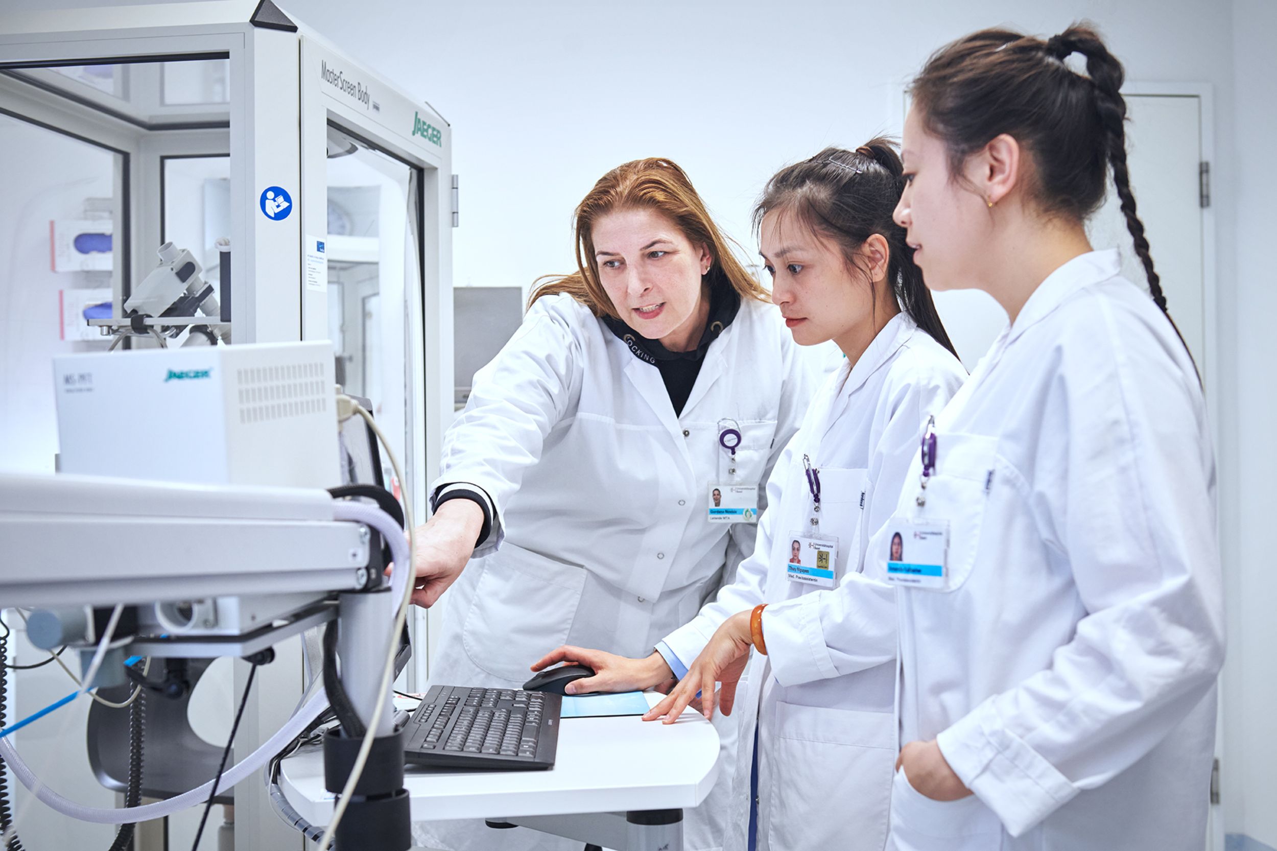
Our pulmonary function laboratory offers all pulmonary function tests on an inpatient and outpatient basis. Breathing tests can be used to examine respiratory performance without any radiation exposure. Deviations from the norm provide important indications of a lung or respiratory disease. Lung function tests can measure the size of the lungs (bodyplethysmography) and the speed of the respiratory burst and respiratory volumes (spirometry). Under certain circumstances, we may stimulate the airways in a further test (bronchoprovocation). If there is a narrowing of the airways, this is seen as an overreaction and is a typical sign of bronchial asthma.
The lung function is supplemented by measuring the gas exchange of the lungs (blood gases, CO diffusion capacity) and determining inflammation values in the exhaled air (eNO). Patients with advanced lung diseases often suffer from a lack of oxygen in the blood, which can be detected with blood gas tests. Oxygen can then be administered at home as a treatment (home oxygen therapy). As lung specialists, we determine the amount of oxygen required for a good oxygen supply (oxygen titration) and carry out regular checks. In some patients, diseases of the muscles or nerves of the respiratory organs are suspected. In this case, the respiratory muscle strength (respiratory mechanics) can be determined.
The above examinations are carried out at rest. Exercise tests must be carried out if the previous tests are not sufficient for the assessment, if we need to find out more about gas exchange under stress or if we want to separate lung problems from heart problems. The simplest examination is the measurement of the walking distance in six minutes (6-minute walk test), the most complex is a so-called spiroergometry, an examination where lung function is performed while cycling and the work of the lungs, heart and muscles is analyzed. This examination is also frequently carried out to determine functional reserves and assess the risk of lung surgery.
Sleep laboratory
Many people suffer from sleep disorders. Breathing-related (respiratory) sleep disorders are a common cause of this. These cause daytime sleepiness and lead to a measurable increase in accidents (microsleeps).
The following examinations are available to clarify sleep disorders:
- Respiratory polygraphy can be used to determine whether sleep and breathing disorders are present using an analyzer similar to a wristwatch, which is worn at home during the night. For further clarification, a nocturnal sleep examination is carried out in the sleep laboratory, which measures not only breathing patterns but also brain waves and many other parameters (polysomnography). Special equipment can be used to measure not only oxygen saturation throughout the night, but also the CO2 present in the blood, which, if elevated, is an indication of respiratory insufficiency.
- Patients with respiratory failure need therapy with nocturnal positive airway pressure (CPAP therapy), which is individually adjusted using a computerized system and must then be carried out permanently at home in the evening. Patients with insufficient respiratory drive or neurological and muscular diseases that affect the respiratory muscles are treated with nocturnal home ventilation. After the start of treatment, the effectiveness is checked at regular intervals and the devices are readjusted if necessary.
Lung endoscopy and pleural surgery
In addition to the clinical examination, laboratory assessment, computer tomograms and lung function, we also carry out bronchoscopies. Patients are asleep during bronchoscopy, but no anesthesia is required. Bronchoscopies are performed for a wide variety of diseases such as lung infections in immunocompromised patients, suspected lung tumors, interstitial diseases, etc. A wide range of technical options are available. Flushing fluid can be taken for bacteria, viruses and fungi.
During lung endoscopy (flexible bronchoscopy), the large airways are viewed using a thin (3-7 mm thick) controllable fiberglass tube (bronchoscope). Bronchoscopy is not painful. Patients are given a sedative for the examination (no general anesthesia) and are therefore unaware of the examination itself. The examination takes about 10-40 minutes. Patients should "sleep in" for a few hours afterwards and are not allowed to drive on the same day. Patients can usually leave the hospital on their own after a maximum of four hours.
A lung endoscopy enables the examination of abnormalities in the lung tissue, such as
- Shadows on the X-ray (infiltrate),
- nodules,
- scarring of the lung tissue (interstitial changes)
- as well as enlargement of the lymph glands (lymph nodes), which are located between the two lungs.
If there are abnormal findings in the lung endoscopy, the lung specialists can flush the lung with a small amount of fluid (bronchoalveolar lavage), scrape the mucous membrane a little or even take a small tissue sample (transbronchial and endobronchial biopsy). Lung endoscopy is also ideal for quickly and painlessly clarifying small lumps next to the airways (fine needle aspiration). With the new technology, endobronchial ultrasound (EBUS), an ultrasound examination can be carried out via the bronchoscope.
With this method, lymph glands located next to the airways can be visualized and a specific sample can be taken from them. Sometimes we observe that the patient's breathing is obstructed by secretions, tissue or small objects in the airways. If this is the case, it can be suctioned out, pulled out with forceps or even reopened with hot argon gas (argon therapy) or high-energy light (laser application). If the airways are severely constricted from the outside, a supporting mesh tube (stent implantation) can be inserted in a few minutes to keep the airway open. The appropriate probes for local post-radiation therapy (brachytherapy) of the airways can also be inserted.
If a disease of the thin fluid-filled space around the lung (pleural space) is suspected, it can be examined as follows:
- first, an ultrasound (pleural ultrasonography) is usually performed. Aspiration of excessive fluid or air from this space (pleural puncture) can bring about a significant improvement in breathlessness or even determine the cause. To do this, a very thin needle or a thin tube must be inserted into this space between the ribs under local anesthesia. The tube often has to remain in place for several days (pleural drainage). In a similar way, a sample can be taken (lung and pleural biopsy) or the chest and lungs can also be examined with a video camera (thoracoscopy). A thoracoscopy is performed on patients who are asleep. This allows us to free an adhered lung (adhesiolysis) and bond partially detached lungs to the chest wall (talc pleurodesis). Patients must be admitted to hospital for a few days for this procedure.
This information does not replace any verbal or written explanation by the doctor. If you have any questions, the doctor on duty or your family doctor will be happy to answer them.
Covid-19
International studies are providing increasing evidence that patients are still suffering from health restrictions even months after they have recovered from Covid-19. The extent of these restrictions has not yet been clarified, so regular check-ups with the family doctor and, if necessary, a specialist are advisable.
As lung specialists, we will be happy to assist you if you continue to have breathing difficulties after contracting Covid-19 that make you feel unsafe. Please contact us on +41 61 265 51 80 or pneumo@usb.ch.
The test center at the University Hospital Basel (USB) was closed on January 31, 2023. You can find other test centers on the website of the Basel-Stadt Department of Health.
Visits and the accompaniment of outpatients are possible. No more than six people - including patients - may stay in a patient room on the wards. One accompanying person is permitted for outpatient stays. Further information can be found under the following link.
The University Hospital Basel and the Basel-Stadt Department of Health provide information on their current assessment of the new coronavirus in the canton of Basel-Stadt. A website of the Department of Health provides helpful information.
