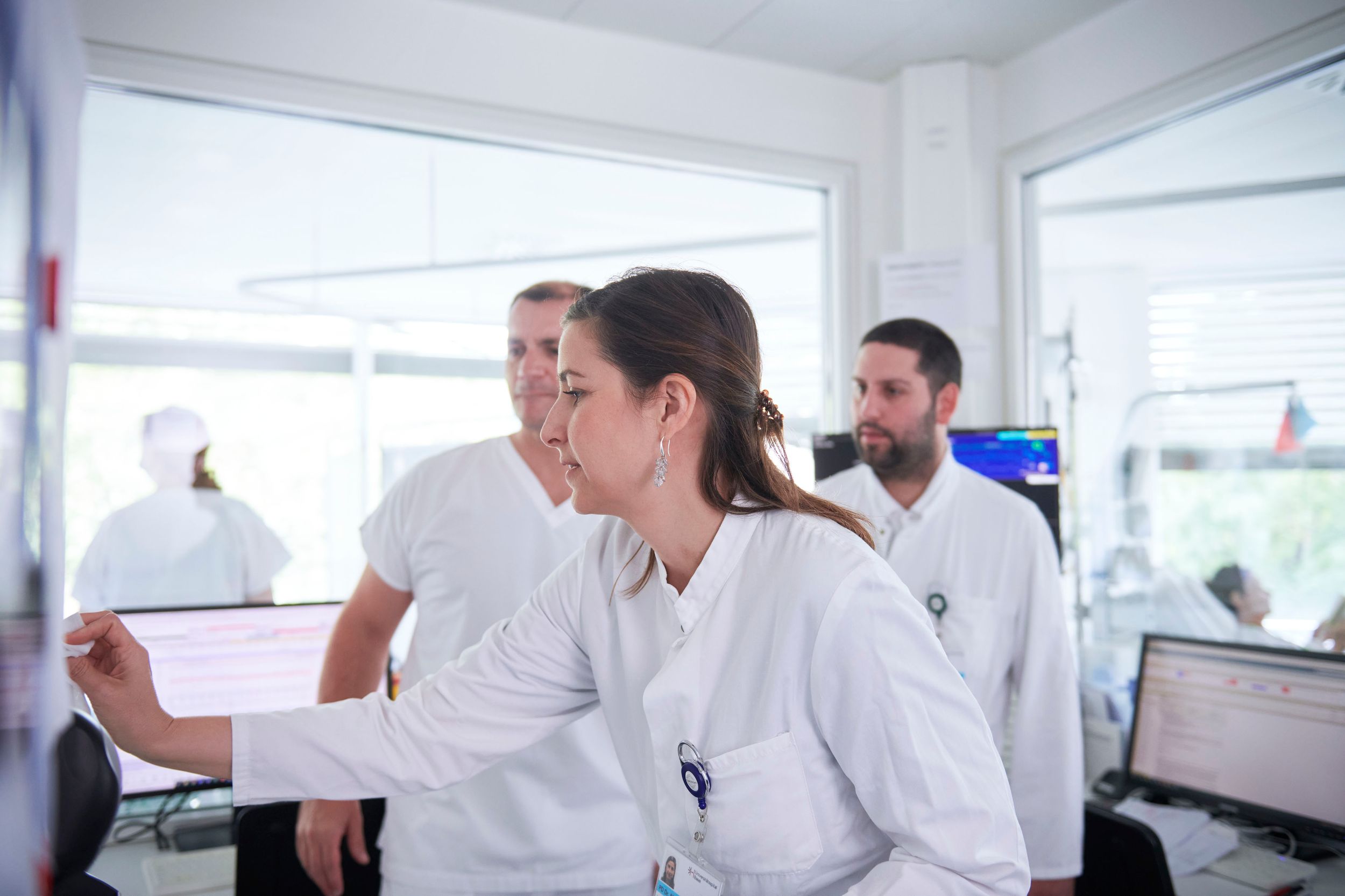
Stroke Center University Hospital Basel - World-leading expertise with a regional commitment
As one of nine certified stroke centers in Switzerland, we offer immediate, comprehensive and quality-tested treatment - for every patient, with the utmost care, expertise and a coordinated assessment and treatment concept.
Our vision is a future in which every stroke patient in Northwestern Switzerland has the best chance of recovery thanks to immediate, first-class and individualized care - locally anchored, internationally pioneering.
Stroke care services - available for you at any time
What is a stroke?
In Switzerland, around 15,000 people suffer a stroke every year. It is the most common cause of permanent disability in adults.
Although the brain is only a relatively small organ, it needs around a quarter of the blood that the heart pumps when at rest. If this blood supply is interrupted by a vascular occlusion, neurological deficits occur after just a few seconds - such as speech disorders, loss of sensation or visual impairment.
There are two main forms of stroke:
- Ischemic stroke
The most common form: Here, the blood supply to an area of the brain is blocked. As a result, the nerve cells receive too little or no oxygen, which quickly damages them.
- Cerebral hemorrhage
In this form, a vessel in the brain bursts. The leaking blood damages the surrounding brain tissue.
Symptoms of a stroke: Recognizing warning signs
A stroke often strikes like a bolt from the blue. The symptoms can occur individually or in combination and vary depending on which area of the brain is affected. However, they are always an alarm signal that requires immediate action. Here are the most important signs:
- (Unilateral) paralysis or numbness
Sudden loss of one side of the body - face, arm or leg. Movements such as lifting an arm become impossible, or there is a feeling of numbness, usually on one side only.
- Visual disturbances
The field of vision changes abruptly: one eye may go blind, vision is restricted to one side, or objects appear double, as if seen through a distorted glass.
- Speech disorders
The ability to speak is lost or severely restricted. Words come out haltingly or not at all, and it is difficult to understand spoken language.
- Spinning vertigo
A persistent feeling as if everything is spinning, accompanied by unsteadiness when standing or walking, often occurs together with other symptoms.
- Headache
A sudden, unusually intense headache sets in that is clearly distinct from normal pain.
Behavior in an emergency
A stroke is a medical emergency in which every minute counts. In the first few hours after the onset of symptoms, it is possible to reopen an occluded vessel and thus prevent permanent damage. The sooner treatment begins, the better the chances of recovery. Early admission to hospital is therefore crucial.
Act quickly with the FAST Check
A stroke can be detected quickly with the FAST symptom check:
Face: Ask the person to smile or show their teeth. Is the mouth crooked or is one corner of the mouth hanging down on one side?
Arm (arms): Ask the person to stretch both arms horizontally forward, raise them and turn their thumbs up. Does one arm drop or stay down?
Speech : Ask the person to speak. Is the speech slurred or difficult to understand?
Time : If one or more of these signs occur, call the emergency number 144 immediately - even if in doubt!
Treatment of stroke - procedure and measures
A stroke requires rapid action, as it poses an acute health risk. At the University Hospital Basel, treatment usually begins in the Emergency Center. The symptoms are recorded there, initial diagnoses are made and immediate measures are initiated. The patient is then admitted to the so-called stroke unit - a specialized ward for patients with an acute stroke.
The path to diagnosis
The Stroke Center starts a comprehensive examination process to find the cause of the stroke and choose the right therapy. The frequently performed steps include
- Recording of visible symptoms, such as paralysis or speech disorders.
- An ECG and ECG monitoring to detect heart rhythm disturbances such as arrhythmias.
- Computed tomography (CT) - with and without contrast medium - to localize circulatory disorders.
- Three-dimensional imaging of the blood vessels to identify constrictions or blockages caused by arteriosclerosis.
- Magnetic resonance imaging (MRI) for detailed insights into the brain.
- Ultrasound examinations of the vascular walls and the heart, for example to detect sources of embolism.
These diagnoses enable the specialists to adapt the treatment individually.
Therapy: time is crucial
Treatment depends on the type of stroke. In the case of a vascular occlusion, an attempt is made to remove it as quickly as possible - either with medication (e.g. clot-dissolving enzymes) or with surgery. The aim is to restore blood flow and minimize brain damage.
Care in the stroke unit at the Stroke Center Basel
The Stroke Unit offers all-round care by a specialized team. This includes
- Continuous monitoring of cardiac and circulatory functions to detect and treat complications at an early stage.
- Nursing care and supportive therapies such as physiotherapy, occupational therapy and speech therapy to promote recovery.
- Research into the causes, for example using ultrasound of the neck and brain vessels or long-term ECGs.
- Measures to prevent further strokes, e.g. through medication or lifestyle changes.
- If necessary, support through social counseling or additional neurological examinations.
Supportive therapies: A key to success
Supportive therapies such as physiotherapy, speech therapy and occupational therapy complement our range of treatments for patients. These measures enable significant long-term progress and make a decisive contribution to improving quality of life. Relatives also benefit from these services, as they support care and recovery.
Occupational therapy
Occupational therapy is used when a person has suffered paralysis after a stroke or has difficulties with mental abilities. This includes, for example, the ability to plan and implement things, to remember or to concentrate. Occupational therapists help those affected to find solutions and aids - for example, how they can use a paralyzed hand in everyday life again. This makes it easier to return to an independent life. Treatment therefore often takes place directly in the patient's room. It can also take place in a therapy kitchen or workshop. The focus there is on analyzing and practicing everyday activities such as household tasks in order to promote independence.
Speech therapy
In the case of speech disorders that manifest themselves in problems with understanding, reading, speaking and writing, the speech therapist tries to find ways to make communication easier. In the further course of treatment, the speech difficulties of those affected are addressed with targeted speech exercises. In the case of speech disorders, which manifest themselves in unclear articulation and changes in the melody of sentences and the sound of the voice, the aim of the therapy is to achieve the best possible intelligibility.
Physiotherapy
After a stroke, patients can be impaired in their independence to varying degrees. Paralysis, changes in sensitivity, perception, coordination and/or movement planning can occur. The event often also results in an impaired ability to walk. The aim of physiotherapy in the acute phase is to re-learn daily movements with the necessary support. The therapist pays attention to the quality of movement in order to avoid learning unnecessary compensatory movements. The physiotherapist's individually adapted and professional assistance is crucial for this. With this support, impaired movements are controlled and the affected side of the body is used again in a targeted manner. Depending on the situation, treatment in the acute phase may require the involvement of two physiotherapists.
Neurorehabilitation
Depending on the severity and course of the illness and the extent of the impairment, further rehabilitation in a specialized institution may be indicated. In this case, the affected patients are transferred to a suitable clinic immediately after their stay with us. At the stroke center, there is a cross-hospital stroke treatment chain to the neurorehabilitation department at Felix Platter Hospital. This is characterized by rotations at ward doctor level and a medical management anchored in both hospitals, which leads to a uniform treatment doctrine. There are also long-standing partnerships with other rehabilitation clinics in the area, which means smooth and unbureaucratic referrals for follow-up treatment.
Risk factors and prevention of a stroke
After a stroke or cerebral haemorrhage, there is an increased risk of recurrence. Targeted aftercare and preventive measures are therefore essential, especially for people with corresponding risk factors. The most common and widespread risk factors include
- High blood pressure
- Untreated heart disease
- Elevated blood lipid levels
- Excessive alcohol consumption
- smoking
- Lack of exercise
- Obesity
- Sleep apnea syndrome
Prevention through specialized advice
In order to promote brain health, we offer comprehensive advice in our specialist consultation hours. These include
- Neurovascular consultation: Neurologists, neurosurgeons and neuroradiologists are available as needed.
- Neurovascular ultrasound: Specialized examinations of the vessels supplying the brain.
- Hypertension consultation: Targeted support for high blood pressure.
- Lipid consultation: Treatment of elevated blood lipid levels.
- Smoking cessation: support in quitting.
- Diagnostic and interventional neuroradiology: CT, MRI and angiography for precise diagnoses.
These services help to minimize risks and sustainably improve quality of life.
Offers for relatives
Social counseling at the University Hospital Basel is an independent specialist group and part of the interdisciplinary treatment team at the hospital. Our self-image is based on a holistic view of physical, psychological and social factors. This means that we work with you and your relatives to identify the respective problem situation and try to develop a viable follow-up solution.
We can advise you on the following topics, among others:
- Outpatient support services
- Exit planning and rehabilitation
- Housing situation and homelessness
- Employment in connection with illness/disability
- Social insurance
- Financial problems and debt situation
- Child and adult protection law
- Addiction disorders
- Migration
- Violence and victim support
If you would like to talk to the social counseling service, please contact your attending physician, the responsible nursing staff or contact us directly. We will be happy to assist you with our professional expertise.
Contact us
Medical Social Services
Thomas Rohrbach
Phone +41 61 265 74 90
Neurorehabilitation and follow-up treatment
Rehabilitation begins as an inpatient in the stroke unit. In severe cases, we offer highly intensive early rehabilitation directly on site.
Lighter cases are treated on an outpatient basis, while inpatient rehabilitation is carried out in cooperation with partners (University Geriatric Medicine Felix Platter, Rheinfelden Rehabilitation Clinic, Cantonal Hospital Baselland / Bruderholz and Liestal, Hôpital du Jura). Our therapy institutes (physiotherapy, occupational therapy, speech therapy) and nursing care are responsible for coordination.
Coordination conference
This conference (at least 6 times a year) optimizes the processes of the stroke unit. Participants: nursing managers, rehabilitation representatives, neurology nursing consultants, medical management of the stroke unit. Depending on the topic, other specialists are consulted.
Partnerships
Information about our partner hospitals:
University Geriatric Medicine Felix Platter: Inter-hospital treatment of stroke patients, from the acute to the rehabilitation phase; neurological co-management of the neurorehabilitation department
Bruderholz: Rehabilitation
Liestal: Multi-hospital treatment of stroke patients, from the acute to the rehabilitation phase
Rheinfelden rehabilitation center: Stroke rehabilitation and neurocognitive assessment and therapy
Hôpital du Jura, Porrentruy site: rehabilitation
REHAB Basel: Rehabilitation of severely affected stroke patients
Quality seal
To optimize the treatment of stroke patients, certified stroke centers have been established in Switzerland, similar to those in some European countries. In spring 2014, the Brain Stroke Center Basel received its official seal of quality as a so-called "Stroke Center" in accordance with the guidelines of the Swiss Stroke Society, the certification requirements of the Swiss Federation of Clinical Neuro-Societies (SFCNS) and the European Stroke Organization (ESO). This quality label distinguishes the Basel Stroke Center as one of the eight certified Swiss Stroke Centers.
We are delighted to have been voted the best specialist clinic in Switzerland in 2024 by the Handelszeitung newspaper.
All common diagnostic methods and treatments for acute cerebrovascular diseases are available on site at a stroke center. This also includes measures that have been assigned to highly specialized medicine. These can be: catheter-based reopening of occluded cerebral arteries in the acute phase or as an elective intervention for secondary prevention, craniectomy, interventional or neurosurgical treatment of intracranial aneurysms and arteriovenous malformations.
The advantages of this benefit our patients: the chance of surviving a stroke and not suffering any disabilities is increased by 25 percent in a specialized stroke center (source: Deutsche Schlaganfall Hilfe) and follow-up treatment is always carried out according to the latest state of knowledge.
