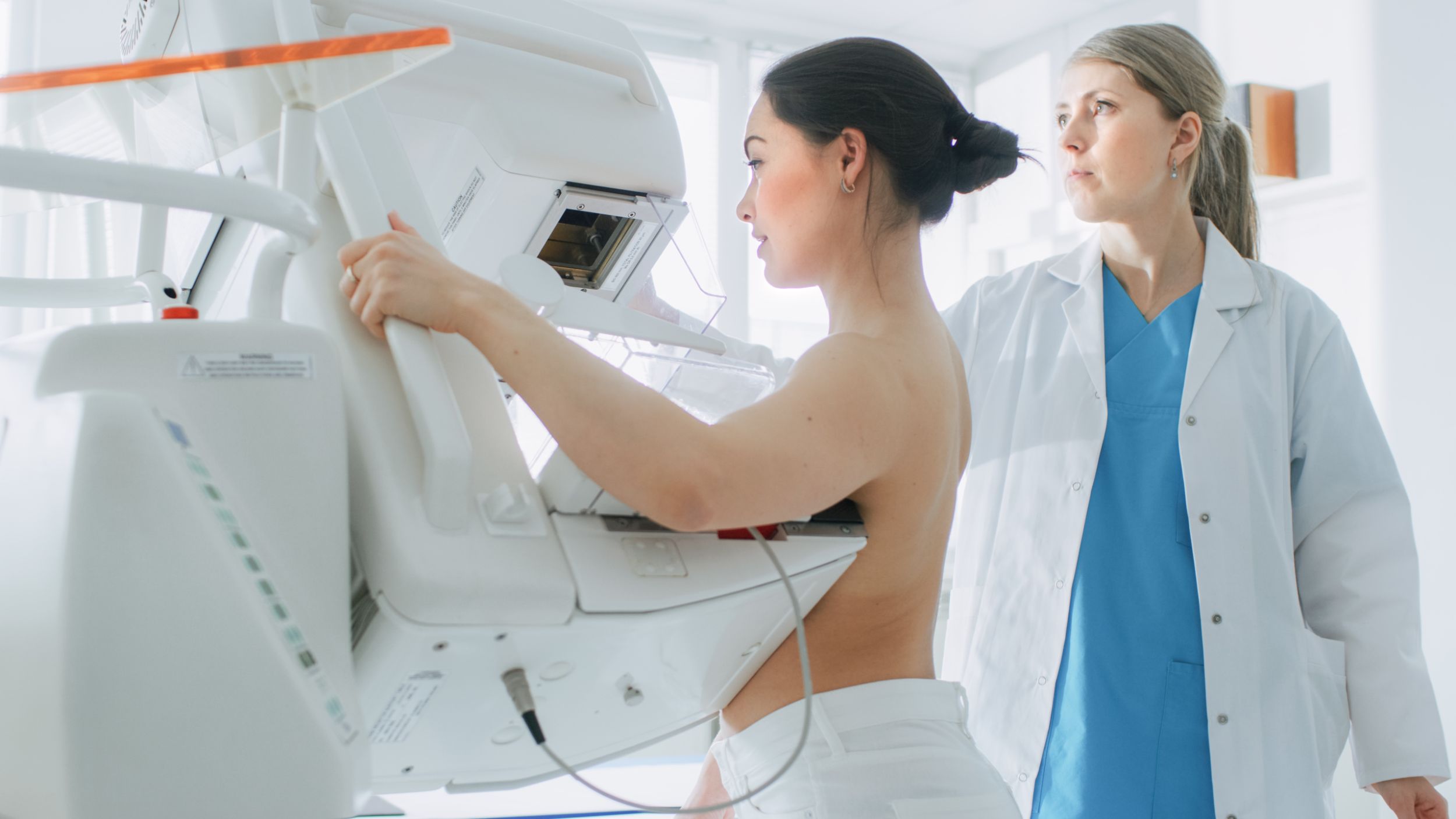
Patient and doctor at the X-ray machine

Offer

On these pages you will find important and useful information so that you can find out about our examinations and therapies. Your well-being is the focus of our work - in the clinic as well as in research and teaching.

Examinations and therapies

Imaging and therapy in figures

300'000

Single images per day

148'000

Examinations and therapies during the year

2'470

Interventions per year

400

terabytes - our image archiving and information system is as extensive as 188,800 hours of television-quality video.

Radiation protection

We attach particular importance to efficient and consistent radiation protection in order to minimize the exposure of our patients and employees to radiation. Responsibility for radiation protection lies primarily in the hands of the Radiological Physics and Radiopharmaceutical Chemistry departments. All departments that work with radiation are actively involved in the implementation of radiation protection.


The tasks of Radiological Physics include the radiation dose monitoring of approx. 700 occupationally exposed persons at the University Hospital Basel, the coordination of quality assurance at the X-ray equipment throughout the hospital, the licensing of the operation of this equipment, dose estimates for X-ray examinations and advice on various radiation protection issues within and outside the hospital.


Radiopharmaceutical Chemistry is responsible for the safe handling of open radiation sources during nuclear medicine examinations and therapies and - in collaboration with the Department of Nuclear Medicine - for the inspection and disposal of radioactive waste.

Dispensing with shielding protective agents during radiological examinations

In the past, lead aprons or other protective agents were used in some radiological examinations to shield organs sensitive to radiation.

Thanks to technical progress, lower radiation doses are now sufficient to obtain good imaging and it is possible to better protect radiation-sensitive organs. This reduces the overall radiation exposure, especially in the direct examination area, and also scatters less radiation into the surrounding organs and body regions.

Due to these developments, we do not use X-ray aprons and other protective devices such as testicular capsules, lens protection or thyroid protection at the University Hospital Basel.

By dispensing with these protective devices, there is also no risk of them unintentionally entering the examination area, which could result in disadvantages such as poorer image quality or a negative impact on radiation exposure.

These statements are based on the latest scientific publications, the recommendations of the Swiss Society for Radiobiology and Medical Physics and the Swiss Federal Commission for Radiological Protection

However, if you feel unsafe or would like additional protection, we will continue to provide you with protective equipment on request.

Please contact us if you have any questions.

Every X-ray and nuclear medicine examination is associated with radiation exposure for the patient. This should be kept as low as possible. However, the corresponding measures to reduce radiation exposure must also be practicable. The most important aspects that help to minimize radiation exposure are

  • a careful indication for the examination
  • high-quality technical equipment
  • good training and experience of the staff

Our clinic meets these and other requirements.

Natural and civilization-related radiation exposure

Humans are exposed to various types of radiation (e.g. light, UV, radio waves, radiation from X-ray equipment and radioactive sources). X-rays belong to the group of ionizing radiation (radiation that can remove electrons from atoms and leave ions behind). When radiation hits the body, some of it is absorbed in the tissue - the amount of radiation absorbed is described by the term dose.

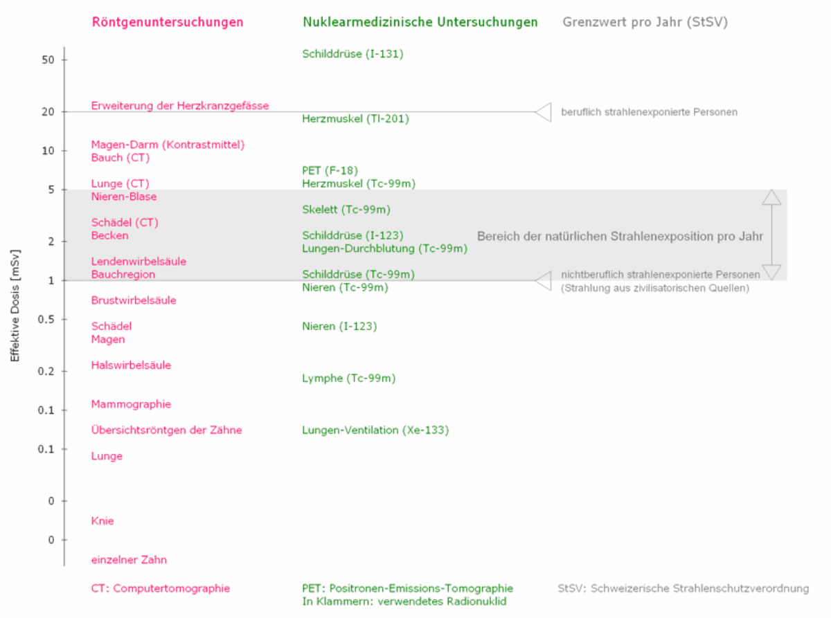
Different radiation exposures can be compared by specifying the so-called effective dose (unit: milli-Sievert, abbreviated to mSv). This provides a measure of the radiation risk to the exposed person. The natural radiation to which all living beings are exposed can be used for comparison. For the Swiss population, the four natural radiation components (cosmic and terrestrial radiation, incorporated radionuclides, radon with by-products) lead to an average effective dose of around 4 mSv/year, whereby the fluctuation range from 1 to around 150 mSv is very large. Civilization's radiation exposure, on the other hand, averages 1.2 mSv/year, almost all of which comes from medicine. Overall, the average radiation exposure of a living being in Switzerland is therefore around 5 mSv/year.

Most X-ray and nuclear medicine examinations, such as images of the lungs, skull and extremities, involve small doses; they belong to the so-called low-dose range. X-rays of the peripheral extremities, including the elbow and upper ankle, are also not considered dose-intensive diagnostic applications.

Dose-intensive X-ray and nuclear medicine examinations

Other examinations count as dose-intensive applications, e.g. X-ray examinations of the axial skeleton (cranial bones, spinal column, pelvis), pelvis and abdomen (stomach) as well as examinations in which several image slices are taken (conventional tomography, computer tomography). Fluoroscopy, fluoroscopy-supported contrast medium examinations and interventions also belong to the group of dose-intensive examinations. In the case of dose-intensive X-ray examinations, the effective dose is generally greater than 0.5 mSv.

Similarly, the effective dose for numerous nuclear medicine examinations is more than 0.5 mSv.

The figures in the following list are typical doses in mSv, which are to be regarded as guide values for the magnitude of the medical radiation exposure in question. Larger individual differences may occur depending on the exposure technique and the constitution, sex and age of the patient.

In contrast to closed radiation sources, such as those used for X-ray examinations, more extensive safety measures are required when handling open radioactive sources due to the risk of contamination.

In order to monitor individual radiation exposure, triage measurements are carried out daily for employees who carry out nuclear medicine examinations and treatments and therefore work with open radiation sources (doctors, laboratory technicians, medical-technical radiology specialists, biomedical analysts, nursing and cleaning staff).

Radioactive waste (e.g. waste water from the ward) must be disposed of responsibly. The Radiopharmaceutical Chemistry and Nuclear Medicine departments are jointly responsible for testing, measuring and disposing of radioactive waste.

Important prerequisites for optimal radiation protection are the safety and proper functioning of X-ray equipment and nuclear medicine devices and accessories. Well-trained specialists, in particular medical-technical radiology specialists (MTRAs), ensure that any malfunctions and deviations are detected in good time.

During commissioning, a quality assurance program checks that the equipment is functioning properly. An acceptance test must be carried out before a medical system or imaging system is used for the first time, followed by periodic constancy tests. Maintenance and subsequent status checks are also carried out regularly. Aids such as lead aprons are checked for their condition at fixed intervals and repaired or replaced if necessary.

At the University Hospital Basel, we naturally carry out the various checks in accordance with radiation protection legislation. We even check individual devices and utensils more frequently than prescribed in order to ensure optimum radiation protection.

Through their behavior, patients can help to ensure that a necessary X-ray examination provides good quality results with as little radiation as possible. Wherever possible, it must be avoided that an examination has to be repeated. Patients can contribute to an optimal X-ray examination by taking the following measures:

  • Follow the instructions of the staff
  • avoid movement during an X-ray (unless instructed to do so)
  • remove jewelry, hair clips, watches, etc. during examinations in the corresponding body area
  • draw the staff's attention to special features (e.g. implants)
  • bring previous X-ray images with you or draw your attention to them
  • Take your X-ray passport with you (if available)

Holding patients in place

Certain patients (e.g. children, frail or elderly patients) may need to be supported or held during the X-ray examination in order to obtain a high-quality X-ray image. Professionally exposed persons (medical-technical radiology specialists, doctors, etc.) may only hold children, unsteady or unstable patients if they cannot be restrained during the X-ray examination and no other persons are available to hold them. Relatives or nursing staff must be called in primarily for this purpose. Persons holding patients must use appropriate protective equipment (aprons).

The radioactive material used at the University Hospital Basel is disposed of responsibly. The Departments of Radiopharmaceutical Chemistry and Nuclear Medicine are responsible for this. We check that waste is stored safely until its radioactivity has decayed and collect radioactive waste water, for example from the nuclear medicine ward (from toilets, washbasins and showers), in separate tanks.


Only when the radiation has reached the limits prescribed by the Federal Office of Public Health (FOPH) do we arrange for the disposal of the now harmless material.


In addition, we take weekly samples of the wastewater from the entire University Hospital Basel and test them for radioactivity.

Contact form Radiology and Nuclear Medicine

Haben Sie eine Frage oder ein Anliegen?

Ihre Meinung ist uns wichtig. Wir schätzen es, wenn Sie mit uns Kontakt aufnehmen und freuen uns über Ihre Anregungen, Hinweise und Kritik. Bitte verwenden Sie dazu unser Formular. Vielen Dank, dass Sie sich dafür Zeit nehmen.

Kontaktinformation