Abdominal and oncological diagnostics (with breast diagnostics)
Specialty and area of responsibility
We deal with the imaging diagnosis of traumatic, acute and chronic diseases of the abdominal and pelvic organs, the female breast (breast diagnostics section) and the minimally invasive diagnosis of blood vessels. We are also dedicated to overarching oncological issues.
At our outstation, the Felix Platter University Department of Geriatric Medicine, we focus on age-related abdominal and oncological diseases.
As experts in abdominal, vascular and oncological imaging as well as imaging care at the Emergency Center, dedicated physicians trained through fellowships are available to provide our patients with modern, high-precision imaging in accordance with current guidelines. Accurate diagnosis is the basis for targeted and effective treatment and is carried out in close cooperation and continuous interdisciplinary exchange (especially in tumor conferences and clinical case discussions/reports) with our specialist clinical colleagues - for optimal patient care.
Fellowships
A fellowship in the Department of Abdominal and Oncological Diagnostics and the Breast Diagnostics Section follows the 5-year training period to obtain the specialist title as a 1-year subspecialization. It can be obtained either at the Department of Radiology and Nuclear Medicine at the University Hospital Basel or at external institutions with an organ-specific focus. The aim of a clinical fellowship is to continue and deepen the training process in a subspecialized discipline: the expansion of specific diagnostic knowledge and the practical consolidation of complex therapeutic procedures. The focus is on assuming responsibility and gaining independence through active participation in the training process of radiology residents, effective interaction with medical management (especially in communicating radiology examination results) and conducting reports.
Clinical and scientific focus
The Department of Abdominal and Oncological Diagnostics is a central partner of the Emergency Center, the Clinics for Internal Medicine, the Medical Polyclinic, the Women's Clinic, Clarunis Gastroenterology and Hepatology, Urology, Clarunis - Visceral Surgery, Vascular Surgery and Clinic for Organ Transplantation, Clinic for Transplantation Immunology and Nephrology, Clinic for Dermatology, Oncology and Pathology.
Our close collaboration with colleagues from these clinics ensures specialist findings, precise diagnoses and individual treatment plans for our joint patients.The increasing complexity of therapeutic procedures requires integrated diagnostics: five times a week, oncologists, surgeons, gastroenterologists, pathologists, radiotherapists and radiologists meet at interdisciplinary and specialist tumor conferences to discuss therapeutic options and determine further measures and therapies.
Breast diagnostics is an integral part of the Breast Center. This specialist area deals with the diagnosis of diseases of the female and male breast.
Clinical focus areas
- MRI of the liver with liver-specific contrast agent to detect the smallest pathological changes in existing liver cirrhosis, healthy tissue or tissue in storage disease, metastases and differential diagnosis of focal liver lesions
- MRI and CT of the pancreas for acute or chronic inflammation, cancer or cystic changes in the pancreas
- MRI of the bile ducts (MRCP) for the diagnosis of stone disease, inflammatory changes and tumor diseases
- MRI of the gastrointestinal tract (HydroMRI) for chronic inflammatory bowel diseases with dynamic MRI to diagnose adhesions in the bowel
- MRI and CT of the kidneys and urinary tract (ureter and bladder). Cross-sectional imaging diagnostics are used for local imaging and, in particular, to detect the spread of tumors of the urogenital organs and to assess the response to treatment
- Multiparametric MRI examination (mpMRI) of the prostate. The MRI images are of great importance if a fusion biopsy is to be performed in addition to the conventional prostate punch biopsy. For the fusion biopsy, suspicious areas are marked on MRI images and targeted by the urologist, which enables a high degree of accuracy even for very small tissue areas that are suspected of being cancerous
- Pelvimetry using MRI (measurement of the birth canal of pregnant patients)
Oncological imaging plays a central role in the diagnosis and treatment of many tumor diseases and is an elementary component of initial diagnoses. Longitudinal oncological-radiological tumor evaluation is essential for the diagnosis of spread (staging) and is integrated into the entire course of the disease by monitoring the response to therapy (response assessment). In the radiological assessment of immuno-oncological therapy, we have the necessary many years of experience in the identification of atypical patterns of response to therapy.
Procedure
Diagnostic imaging in the Department of Abdominal and Oncological Diagnostics is based on computed tomography (CT), magnetic resonance imaging (MRI), sonography (ultrasound) and conventional X-ray diagnostics as part of fluoroscopy-guided examinations of the gastrointestinal tract. Minimally invasive blood vessel diagnostics are carried out using CT and MR angiography and non-invasive Doppler sonography.
In computed tomography, we focus on radiation dose reduction, together with research groups at the University Hospital Basel (oncology, urology), other university hospitals (Duke University, University Hospital Zurich, University Hospital Bern) and industrial partners (Siemens Healthineers).
The Diagnostic and Interventional Mammography Section uses digital mammography and tomosynthesis methods, including contrast-enhanced mammography, high-resolution breast ultrasound andbreast ultrasound and breast MRI, both for patients who have already had breast cancer and for patients with a significantly increased risk of breast cancer as part of the intensified early detection (screening) and aftercare program.
The PET/CT oncology and nuclear medicine procedure is carried out on an interdisciplinary basis with the Department of Nuclear Medicine at the Department of Radiology and Nuclear Medicine at the University Hospital.
Abdominal and oncological diagnostics team
Department management

Prof. Daniel Boll
Stv. Chefarzt Radiologie und Nuklearmedizin
Leitung abdominelle und onkologische Diagnostik, med. Dienstleistung
Tel. +41 61 328 63 84
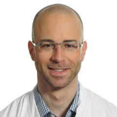
Prof. Dr. Tobias Heye
Leitender Arzt
Leitung Informationstechnologie, Stv. Leitung abdominelle und onkologische Diagnostik
Tel. +41 61 328 63 24
Head of breast diagnostics section

Dr. Noemi Schmidt - EBBI
Leitung Mammadiagnostik, Kaderärztin, Mitglied Tumorzentrum
Radiologie und Nuklearmedizin
Tel. +41 61 265 91 50

Dr. Dilyana Fricker - EBBI
Stv. Leiterin Mammadiagnostik, Oberärztin, Mitglied Tumorzentrum
Radiologie und Nuklearmedizin
Tel. +41 61 265 91 50
Management physicians
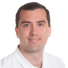
Dr. Konrad Appelt - EBIR und EBBI
Kaderarzt interventionelle Radiologie und Mammadiagnostik
Radiologie und Nuklearmedizin
Tel. +41 61 328 56 31
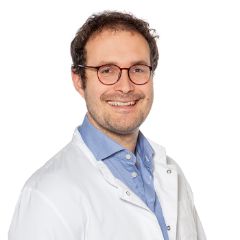
Dr. Hanns-Christian Breit - Dipl. Phys.
Kaderarzt abdominelle und onkologische Diagnostik, Leiter Forschungskoordination
Radiologie und Nuklearmedizin
Tel. +41 61 328 56 33

Verena Funk
Kaderärztin abdominelle und onkologische sowie muskuloskelettale Diagnostik, Leitung Sonografie
Radiologie und Nuklearmedizin
Tel. +41 61 328 73 71
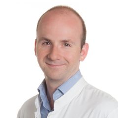
PD Dr. Markus Obmann
Kaderarzt muskuloskelettale sowie abdominelle und onkologische Diagnostik, Leitung Computertomografie und Röntgendiagnostik, stv. Leitung muskuloskelettale Diagnostik
Radiologie und Nuklearmedizin
Tel. +41 61 556 59 05

PD Dr. Jan Vosshenrich
Kaderarzt abdominelle und onkologische sowie muskuloskelettale Diagnostik
Radiologie und Nuklearmedizin
Tel. +41 61 328 51 15
Senior physicians
Pia Brandt
Stv. Oberärztin abdominelle und onkologische Diagnostik
Radiologie und Nuklearmedizin
Tel. +41 61 328 54 06

Dr. Merve Erdogan
Stv. Oberärztin Mammadiagnostik
Radiologie und Nuklearmedizin
Tel. +41 61 328 42 77

Dr. Dilyana Fricker - EBBI
Stv. Leiterin Mammadiagnostik, Oberärztin, Mitglied Tumorzentrum
Radiologie und Nuklearmedizin
Tel. +41 61 265 91 50
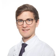
Dr. Moritz Neubauer - EDGAR
Oberarzt abdominelle und onkologische Diagnostik
Radiologie und Nuklearmedizin
Tel. +41 61 328 63 52

Alexander Titov
Stv. Oberarzt und Fellow abdominelle und onkologische Diangostik
Radiologie und Nuklearmedizin
Tel. +41 61 328 42 58
Fellows


Alexander Titov
Stv. Oberarzt und Fellow abdominelle und onkologische Diangostik
Radiologie und Nuklearmedizin
Tel. +41 61 328 42 58
Research assistants
Assistance

Suzana Mehmedi
Assistentin Radiologie, Datenmanagement Radiologie
Radiologie und Nuklearmedizin
Tel. +41 61 328 47 39
