
Teaching
University teaching
The Department of Radiology and Nuclear Medicine is closely linked to the University of Basel(Faculty of Medicine, Departments of Cross-Sectional Medicine and Basic Medical Sciences). Our courses form an essential part of the human medicine and dentistry courses. They are practice-oriented, but also cover the entire spectrum of methods, including complex technologies. In clinical teaching, we offer concept lectures, which are supplemented by practical image interpretation courses.
In addition, our natural scientists in radiological physics conduct various lecture series and seminars for the Biomedical Engineering Master's program of the Department of Biomedical Engineering.
Practical training
We offer internships and work shadowing in a quality-conscious company with state-of-the-art technical equipment and a wide range of examinations:
- for students of human medicine and dentistry in their elective year of study
- for prospective radiology specialists HF (MTRAs)
- for students on the Business Administration-Healthcare Management course
Further education and training
We attach great importance to the further development of our employees' expertise. We are therefore committed to both theoretical and practical training. In terms of further medical training, our clinic meets the highest requirements of the Swiss further training regulations (category A) and is therefore authorized to provide full further training as a specialist in radiology or nuclear medicine. In addition, we offer in-depth training in the specialist areas of radiology through regular rotations and long-term fellowship positions in our specialist departments. A special feature is the organ-based structure of our fellowship program. Doctors and radiology specialists benefit from our regular internal training events and are supported by us in attending external events, congresses and courses.
Medical studies
Radiology and nuclear medicine are method-oriented subjects with concrete clinical relevance. Lectures and practice-oriented, case-based courses provide students with basic knowledge of diagnostic and therapeutic radiology and nuclear medicine. The subjects play a central role in the diagnosis, treatment planning and follow-up of diseases and trauma.
In accordance with the longitudinal structure, the application and technical principles of the procedures as well as the radioanatomy of healthy structures are demonstrated in the Bachelor's program. Students are familiarized with the basics of radiation physics and radiation protection measures in order to ensure the appropriate indication and safe application of the procedures.
Clinical applications and indications, radiomorphology of pathological findings, image interpretation and differential diagnostics as well as treatment options and monitoring follow in the Master's program. The use of radiological data as biomarkers to quantify tissue changes and predict disease progression as well as support from artificial intelligence are also covered.
Basic knowledge of imaging, image-guided and interventional procedures is taught: X-ray, computed tomography - CT, magnetic resonance imaging - MRI, ultrasound/sonography, angiography.
In the courses on nuclear medicine, students learn about the use of radioactive substances for diagnosis and treatment (radionuclide therapy). They are introduced to the interpretation of nuclear medicine images and learn how the techniques (positron emission tomography - PET), single-photon emission computed tomography - SPECT and scintigraphy) are used in combination with computer tomography to visualize molecular structures, analyze organ functions and metabolic processes, as well as for therapeutic applications.
Specialists responsible for the Human Medicine and Dentistry curricula (for Radiology and Nuclear Medicine)
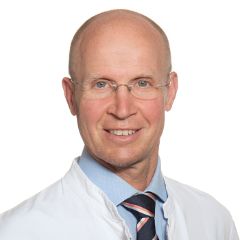
Prof. Christoph J. Zech
Leitung interventionelle Radiologie
Radiologie und Nuklearmedizin
Leitung Angiografie
Tel. +41 61 328 63 44
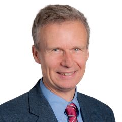
Prof. Dr. phil. Dr. med Damian Wild
Leitung Nuklearmedizin
Radiologie und Nuklearmedizin
Radiologie und Nuklearmedizin
Tel. +41 61 556 53 84
Teaching coordination
Dr. phil. Seline Schellenberg
Wissenschaftliche Mitarbeiterin (Lehrkoordination, Öffentlichkeitsarbeit)
Radiologie und Nuklearmedizin
Structure and content
Human Medicine
Radiology and Nuclear Medicine are involved in all annual courses of the Bachelor's and Master's curriculum in Human Medicine at the University of Basel and in almost all subject blocks.
In the first year of the Bachelor's program, our natural scientists teach the basics of both physics and chemistry, the latter with the support of the Department of Chemistry of the Faculty of Natural Sciences. We also offer a practice-oriented introduction to the technology and application of radiological procedures.
Inthe 2nd and 3rd Bachelor's year, the focus is on radioanatomy and what has been learned is deepened in the subject blocks, a compulsory anatomy module and doctor-patient lessons.
Our training model consisting of concept lectures and assigned practice-oriented differential diagnostic image interpretation courses is part of the program in the 1st and 2nd Master's years.
From the 1st Bachelor's to the 3rd Master's year, we offer practical general radiology as well as specific neuroradiology and interventional professional field studies and elective projects (sometimes for students of human medicine and the medical-technical radiology HF course). Elective projects also take place with our external lecturers in their clinics and thus provide an insight into various radiology departments.
In addition to our courses for human medicine, we also participate in the dental medicine curriculum - with specifically adapted courses on radiology, radiation physics and protection, and nuclear medicine.
Teaching methods and resources
Radiology and nuclear medicine are at the interface between pathomorphology and the clinic. A systematic understanding of pathomorphological changes and the conclusions that can be drawn about underlying diseases are crucial for the correct interpretation of radiological and nuclear medicine examinations.
The concrete application of this knowledge takes place in courses that are integrated into the relevant subject blocks on an organ-specific basis. Here, representative cases are presented in the sense of 'problem-based learning', which are first interpreted independently in small groups and then systematically analyzed together.
Our recommendations on teaching materials have also been integrated into the recommendations of the Faculty of Medicine. In addition, all didactically useful documents for our courses are available online to students for in-depth self-study via the University of Basel platforms.
Our documents are protected by copyright. All rights are held by the Department of Radiology and Nuclear Medicine at the University Hospital Basel. Reproduction and further use of the illustrations and texts is only permitted with the consent of the authors.
Junior assistant, clinical traineeship and internship
Sub-assistance
We will introduce you to the fields of radiology and nuclear medicine by immersing you in our day-to-day work. You will take part in every step of the process, from determining the indication to weighing up the risks and benefits, planning, performing, evaluating and reporting examinations. Depending on the length of your stay with us, you will become familiar with the entire spectrum of our examination equipment as well as complex post-processing algorithms.
We are not always close to the patient (with the exception of nuclear medicine therapies and interventional and neuroradiology procedures), but we are close to the diagnosis and key clinical decisions. Thanks to our organ-specialized organization, we are able to obtain particularly comprehensive, relevant information and incorporate it into clinical discussions and decision-making in a targeted manner. Based on typical pathologies, you will get to know and appreciate the importance of modern diagnostics. The broad medical spectrum covered in our clinic is a great advantage and enables you to significantly expand your pathomorphological understanding of diseases.
You will be able to deepen your knowledge considerably by participating in and contributing to our further and advanced training courses. Depending on the length of your stay (at least 1 month, rotations after 2 weeks), you will rotate to abdominal and oncological diagnostics, cardiothoracic diagnostics, musculoskeletal diagnostics and neuroradiology, or, if you are specifically interested, to nuclear medicine or interventional radiology.
Learning objectives
The professional learning objectives include expanding your knowledge of the appropriate use of radiological methods and the qualified interpretation of examination results as well as the pathomorphological understanding of diseases:
- Checking the indication against the background of medical history, clinical questions and given (preliminary) findings
- Weighing up the benefits and risks of each examination
- Monitoring of examinations and quality assurance
- Systematic analysis of pathological changes with differential diagnostic assessment in the clinical context
- Case presentations at clinical reports, conferences
- Participation in emergency care
Active participation in the daily work of the various specialist areas and departments will give you an overview of the entire field of radiology and nuclear medicine (depending on the rotation).
Tasks
Your tasks will include
- familiarization with radiological and nuclear medicine methods (as well as technical procedures)
- (under supervision) independent diagnosis of conventional chest and skeletal X-rays
- (under supervision) Independent performance and diagnosis of sonographies
- Participation in the diagnosis of cross-sectional imaging examinations (CT, MRI, if interested also SPECT/CT and PET/CT) with post-processing, image analysis and preparation of findings
- Participation in clinical reports and conferences
- Active participation and collaboration in our further education and training courses (e.g. case presentations in our internal lunchtime reports)
Requirements and availability
Students of our faculty in their elective year of study as well as students in their final year of study (for foreign students) are very welcome. You should have a detective-like interest in diagnostic imaging.
The following literature is to be acquired before the start of the sub-assistance:
M. Reiser, F.-P. Kuhn, J. Debus: Radiologie, Thieme, 20174 (esp. chapters "Rad. Procedures", "Nuclear Medicine", "Skeleton"). For further literature, see our teaching material recommendations.
Available places: 1-4/month
Duration and choice of work area
Program duration: 1-4 months with full workload, mandatory from the first day of the month. Depending on the length of stay, the following areas of work are possible:
- Radiology: rotations are possible in abdominal and oncological diagnostics, cardiothoracic diagnostics, musculoskeletal diagnostics and neuroradiology (minimum duration: 1 month, rotation change after 2 weeks in each case).
- Nuclear medicine (on request, minimum duration: 1 month)
- Interventional radiology (on request)
We take preferences into account where possible, but cannot guarantee a desired assignment.
Application modalities
Please send your application with the following documents in digital form to Yasmine Wick
- Letter of motivation
- CV (preferably with photo)
- Current proof of enrollment
- Examination documents (certificates of previous academic achievements)
- Copy of ID (for students from abroad)
- Please specify in which specialty you would like to work: Radiology, Nuclear Medicine, Interventional Radiology).
You can submit your application at any time. Ms. Yasmine Wick (application procedure) and the Radiology and Nuclear Medicine secretariat will be happy to answer any questions you may have.
Clinical traineeships and internships
The same conditions apply to clinical traineeships as to internships. However, they are unpaid.
There is also the possibility of getting a taste of what it is like to work with us for a shorter period (1-3 weeks) as part of a work shadowing program.
Please also contact Yasmine Wick and the Radiology and Nuclear Medicine secretariat .
Examinations
The assessment of learning success in radiology and nuclear medicine is carried out in three stages:
- Examination of the knowledge acquired in the MC (multiple choice) examinations of the individual annual courses
- Examination of the acquired skills of correct image interpretation as part of the OSCE examinations (Objective Structured Clinical Examination)
- Examination of the overall knowledge acquired in the federal examination (human medicine or dentistry)
The content and learning objectives of the individual radiology and nuclear medicine courses can be found in the respective course catalogs. Further information on the MC and OSCE examinations can be found on the Faculty of Medicine website. The Office of the Dean of Studies can also provide information.
Radiology examination as part of the federal examination (human medicine or dentistry)
The latest information on the federal examination in human medicine and dentistry can be found on the website of the Federal Office of Public Health.
Master theses
We supervise students writing Master's theses at the Faculty of Medicine and the Faculty of Natural Sciences.
Interested students can find suggestions via the Dean's Office Master's Thesis Exchange, but can also contact us directly with suggested topics:
Dr. phil. Seline Schellenberg
Further training as a specialist in radiology
You can also find more information:
- on the page Working with us - as a doctor
- in the article "Excellent further training", which appeared in our 2019 annual report. The article is based on an interview with our junior doctor Dr. Moritz Vogt.
Our further training concept is aimed at doctors who want to continue their training in an inspiring environment. The success of our program is based on
people
- Dedicated junior doctors who want to get to know, understand and help shape the fascinating world of radiology
- Renowned experts who pass on their knowledge and enthusiasm for radiology
culture
- Everyone is encouraged to get involved and develop our clinic further.
- By participative leadership, we mean involving employees in the decisions that affect them.
- Clear and transparent rules that make sense for everyone
Organization
- Systematic rotation through all departments
- Possibility of subspecialization
Contents
- Consistent implementation of subspecialized organ-based radiology in accordance with the guidelines of the European Society of Radiology
- Radiology is multifaceted, and soft skills such as leadership, management and economics are also important to us.
Infrastructure
- Our technical equipment meets the latest standards.
- Our diagnostic rooms are organized according to organ departments so that we can easily exchange information and learn from each other.
Innovation
- The continuous improvement of our clinical processes is a matter of course for us.
- Our research topics are relevant, up to date and clearly structured.
Quality
- Our program is certified by the European Board of Radiology and has been awarded the Certificate of Excellence in the best category.
- Our program is certified by the Swiss Institute for Continuing Education and Training
- We received the SIWF Award 2019: Former residents who were supervised as part of their further training in Basel had nominated our further training team for this award for special commitment and competence in continuing medical education.
Does our training concept appeal to you? Then we look forward to receiving your application with a letter of motivation, CV and certificates (preferably by e-mail with PDF).
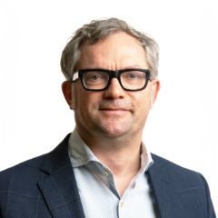
Prof. Jens Bremerich
Leitung kardiale und thorakale Diagnostik
Leitung Aus-, Weiter- und Fortbildung, Radiologie und Nuklearmedizin
Tel. +41 61 328 63 44
Fellowship in Radiology
The aim of a clinical fellowship with us is to deepen further training in a sub-specialized discipline in order to expand specific diagnostic knowledge and to deepen practical knowledge of complex therapeutic procedures.
Responsibility and independence should be assumed within the framework of the:
- active participation in the further training of radiology residents, including organizational responsibility within the department
- effective interaction with the medical management, in particular in the final communication of radiological examination results
- Implementation of reports
To successfully complete the fellowship, comprehensive expertise in the following areas is required: specific professional competence, effective communication, ability to work in a team, efficient self-organization, participation in teaching, basic understanding of professional policy, professional conduct.
We offer clinical specializations/fellowships in the following subspecialties:
- abdominal and oncological diagnostics
- mammography
- Cardiac and thoracic diagnostics
- musculoskeletal diagnostics
- interventional radiology
- Diagnostic and interventional neuroradiology
Prerequisites
- Completed or almost completed further training in radiology
- Specialist diploma recognized in Switzerland
- German language skills at level C1
If you are interested and meet the requirements, we look forward to receiving your application with the following documents (as PDF):
- Letter of motivation
- curriculum vitae
- certificates
Petra Clavette
Assistentin Chefarzt und Teamleiterin der Sekretariate sowie des Datenmanagements
Radiologie und Nuklearmedizin
Tel. +41 61 328 63 84
Further training as a specialist in nuclear medicine
You can find more information in our further training concept for specialists in nuclear medicine.
Does our training concept appeal to you? Then we look forward to receiving your application with a letter of motivation, CV and certificates (preferably by e-mail in PDF format).
Prof. Dr. phil. Dr. med Damian Wild
Leitung Nuklearmedizin
Radiologie und Nuklearmedizin
Radiologie und Nuklearmedizin
Tel. +41 61 556 53 84
Training as a radiology specialist
We train radiology specialists as part of their practical training. This enables them to obtain a diploma as a radiology specialist HF.
The federally recognized training course is carried out at the higher technical college (HF) level. We offer this course together with the Basel-Stadt Health Education Center (BZG). We work closely with the BZG and maintain a regular exchange of information. Further information, especially on the theoretical part of the course, can be found on the BZG website. www.bzgbs.ch
You can find more information about practical training as a radiology specialist at the Clinic for Radiology and Nuclear Medicine via the navigation bar, and the following video of our students will give you an initial impression.
Content and structure of the training
Training to become a radiology specialist takes three years.
It is divided into theoretical sequences at the Basel-Stadt Health Education Center (BZG) in Münchenstein and practical sequences at the training location, for example the University Hospital Basel:
After successfully completing the course, students receive the federally recognized diploma as a radiology specialist HF.
Content of the practical training
The practical training sequences take place in all specialist areas of radiology (diagnostic radiology, radiation oncology and nuclear medicine). We use the following procedures and forms of therapy at the Clinic for Radiology and Nuclear Medicine:
- conventional X-ray diagnostics
- fluoroscopy
- mammography
- computer tomography
- Magnetic resonance imaging
- angiography
- Scintigraphy (static, dynamic, perfusion and receptor scintigraphy)
- Positron emission tomography/computed tomography (PET/CT)
- Single photon emission computed tomography/computed tomography (SPECT/CT)
- Endocrine diagnostics
- osteodensitometry
- Radionuclide therapy (tumor, joint and pain therapy)
- Production of radiopharmaceuticals (radiological chemistry)
For examination procedures and forms of therapy in radiation oncology and radiology in pediatrics and adolescent medicine, see the following websites:
Part of our training concept is that the students complete an external internship planned by us and thus expand their training spectrum.
Training salary
The financial remuneration of around CHF 50,000 starts on the first day of training and is broken down as follows:
- CHF 1100 per month in the first year of training
- CHF 1,300 per month in the second year of training
- CHF 1,500 per month in the third year of training
Possibility of a specialized baccalaureate
During the course of training to become a qualified radiology specialist HF, graduates of the Fachmaturitätsschule (FMS) have the option of submitting their specialist baccalaureate thesis in the field of health and thus obtaining the specialist baccalaureate.
Prerequisites
Persons with the following previous education are admitted to training as a qualified radiology specialist HF:
- Completion of FMS (specialized baccalaureate school) with or without specialized baccalaureate
- High school diploma
- Graduation from a commercial secondary school
- Graduation from the Rudolf Steiner School with IMS-F certificate
- Federal certificate of proficiency (e.g. healthcare assistant - FAGE) with or without vocational baccalaureate
Applicants should also meet the following requirements:
- Interest and sound knowledge in scientific subjects such as physics, chemistry and biology
- Technical understanding
- initiative, organizational and improvisational skills
- good powers of observation
- Accurate and careful work
- communication skills
- ability to work in a team
- Empathy and patience
- Reliability and a sense of responsibility
Apprenticeships
Our students have access to an excellent infrastructure as part of their training to become qualified radiology specialists. As the largest training center for radiology specialists in Northwestern Switzerland, the Department of Radiology and Nuclear Medicine - together with the Department of Radiotherapy and Radiation Oncology - has excellent imaging and therapeutic equipment as well as extensive experience in the training and education of its specialist staff.
Training starts twice a year, in the spring and fall semesters; we offer six students a training place each time.
Further information: The Bildungszentrum Gesundheit Basel-Stadt (BZG) organizes monthly information events on the training course. The current dates can be found on the BZG website or on request
BZG Health Education Center Basel-Stadt
Binningerstrasse 2
CH-4142 Münchenstein
Phone +41 (0)61 417 77 77 | Fax +41 (0)61 417 77 78
bzg@bzgbs.ch | www.bzgbs.ch
If you have any questions or queries, please do not hesitate to contact us personally:
Application
You can apply electronically for one of our training places via the application platform https://www.abhf.ch. Select the University Hospital Basel and upload your documents. If they meet the requirements, we will be happy to invite you to an interview, where we will get a personal impression of you - and you of us. You will also have the opportunity to clarify any open questions directly.
Aptitude internship
The internship lasts three days. As a rule, we will inform you of the result in writing within 14 days. If you pass the internship, we will forward your dossier to the training provider for review.
Admission to training
The admissions committee examines all the documents and decides on admission to training at the BZG.
Once the admissions committee has made a positive decision, we - as the practice institution - can conclude the contract with you. Please do not hesitate to contact our Vocational Training Officer, Georgia Kolakovic, to clarify any questions you may have.
BMA training
With us you can complete the practical part of the training required for the diploma as a biomedical analyst HF (BMA). We offer this together with the Bildungszentrum Gesundheit Basel-Stadt (BZG). We work closely with the BZG and maintain a regular exchange of information. Further information, especially on the theoretical part of the training, can be found on the BZG website.
We have been training students to become BMAs for around 25 years. Every year, we supervise a student during her six-month internship. During this time, she gets to know a large part of our routine work and writes a dissertation documenting her training with us.
We would be happy to inform you personally:
Daniela Biondo Carluccio
Ausbildungsverantwortliche der BMAs
Radiologie und Nuklearmedizin
Tel. +41 61 556 57 22
RapMed e-learning platform
Lecturers in radiology and former students of human medicine at the University of Basel have developed the electronic learning platform RapMed, which can be used to teach radiology and nuclear medicine content on a case-based basis.
RapMed allows lecturers to quickly and easily upload medical cases and students to work out solutions together, expand and test their knowledge, and learn both alone and in groups, in discourse.
The case-based solutions are worked out together. First, the users mark the pathologies. A heat map then shows the selected solutions that stand out most clearly according to their frequency. This graphic labeling is supplemented by a comment function. Through use, the interpretations become more and more accurate - and the platform better and better.
We hope you enjoy it and look forward to your feedback. This is very important, as RapMed is designed to be further developed with the help of feedback from students and lecturers.
Maurice Henkel, Department of Radiology and Nuclear Medicine, University Hospital Basel
RapRad e-learning platform
RapRad aims to enable students of human medicine and assistant doctors to practice the interpretation of medical images so that they can recognize not only the clinical pictures they encounter in everyday clinical practice, but all important pathologies.
The platform, which was awarded the 2018 Lecturer Prize of the University of Basel's Faculty of Medicine, is structured like a computer game - the interpretation of radiological images is taught in a playful way through fast, case-based learning and direct feedback.
RapRad was developed by the Department of Radiology and Nuclear Medicine in collaboration with the Business Informatics department at the FHNW (University of Applied Sciences and Arts Northwestern Switzerland).
We wish you lots of fun and look forward to your feedback(raprad@usb.ch).
Tobias Heye, Philipp Brantner and David Winkel
Department of Radiology and Nuclear Medicine, University Hospital Basel